![]()
![]()
![]()
![]()
2012-06-30

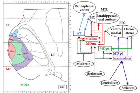
The functional role of the mediodorsal thalamic nucleus (MD) and its cortical network for recognition and memory recall is discussed controversially. Neuropsychologists and Biopsychologists from Bochum now decided to take a fresh approach: They studied patients with focal thalamic lesions to examine the functions of the MD, the intralaminar nuclei and the midline nuclei in memory processing. In addition, a newly designed pair association task was used, which allowed the assessment of recognition and cued recall performance. Most importantly, volume loss in thalamic nuclei was estimated with a technique that had been invented in animal research but was used in this study for the first time on humans. These quantitative anatomical data could then be used as a predictor for alterations in memory performance. The results reveal that reduced recall of picture pairs and increased response times during recognition followed by cued recall covaried with the volume loss in the parvocellular MD. This pattern suggests a role of this thalamic region in recall and thus recollection, which does not fit into the classic framework that is in use in today's Neuropsychology. The functional specialization of the parvocellular MD accords with its connectivity to the dorsolateral PFC, highlighting the role of this thalamocortical network in explicit memory.
The functional role of the mediodorsal thalamic nucleus (MD) and its cortical network for recognition and memory recall is discussed controversially. Neuropsychologists and Biopsychologists from Bochum now decided to take a fresh approach: They studied patients with focal thalamic lesions to examine the functions of the MD, the intralaminar nuclei and the midline nuclei in memory processing. In addition, a newly designed pair association task was used, which allowed the assessment of recognition and cued recall performance. Most importantly, volume loss in thalamic nuclei was estimated with a technique that had been invented in animal research but was used in this study for the first time on humans. These quantitative anatomical data could then be used as a predictor for alterations in memory performance. The results reveal that reduced recall of picture pairs and increased response times during recognition followed by cued recall covaried with the volume loss in the parvocellular MD. This pattern suggests a role of this thalamic region in recall and thus recollection, which does not fit into the classic framework that is in use in today's Neuropsychology. The functional specialization of the parvocellular MD accords with its connectivity to the dorsolateral PFC, highlighting the role of this thalamocortical network in explicit memory.
Copyright © BioPsy 2023
Last update: Oct 04, 2023