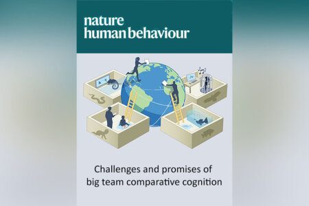

Big team science has the potential to reshape comparative cognition research, but its implementation — especially in making fair comparisons between species, handling multisite variation and reaching researcher consensus — poses daunting challenges. In this paper, a group of authors propose solutions and discuss how big team science can transform the field.
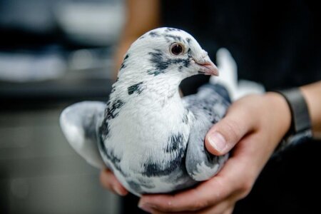

The saying “context is everything”. Quotes, events, actions, or stimuli, cannot be viewed in isolation. They must be interpreted in the light of a bigger picture – their context. This is also evident in extinction learning where, in contextual renewal, an extinguished response reoccurs if the context is changed.
But what exactly is context? In our study, we assessed how competing stimuli induced contextual renewal during extinction learning in pigeons (Columba livia) using a modified ABA paradigm (see below). Manipulating the timing of stimuli, we found that with the right contiguity, small local stimuli resulted in the strongest contextual renewal.
This result challenges definitions of context as ‘a backdrop where learning occurs’. We rather propose that context can be understood mechanistically as a learned stimulus property.
Press release from Science Magazine: >>
Direct link for the publication: >>
ABA Paradigm:
A: The pigeons learn stimulus associations under local and global context factors.
B: Extinction of learned associations under new local and global context factors.
A’: In the renewal phase, one of the two possible context stimuli (local/global) is now presented in its original configuration before extinction , while the other stimulus remains as it was during extinction. This “competition” between the different contextual stimuli enables a comparison of the contextual renewal between the different types of contexts.
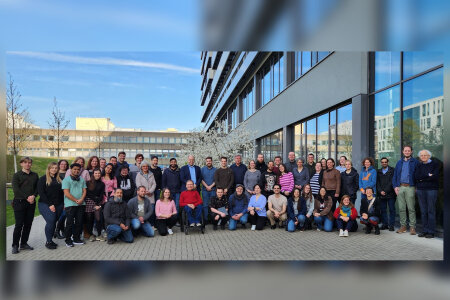

From March 31 to April 2, 2025, we had the pleasure of welcoming around 50 researchers from the SFB 1372 network to our Biopsychology Group at Ruhr University Bochum. Colleagues from Oldenburg, Wilhelmshaven, and Berlin joined us for three days of lively scientific exchange, new ideas, and collaborative planning.
The program included a range of presentations, poster sessions, and informal discussions in smaller groups. In addition, we invited our guests on a guided tour through our labs and shared some spring sunshine during walks through the university’s botanical garden. We would like to thank all participants for their visit and the inspiring conversations – it was a great pleasure to host you in Bochum!
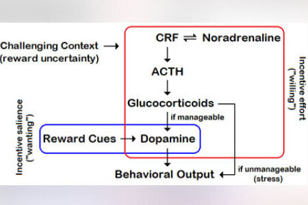

Incentive salience theory both explains the directional component of motivation (in terms of cue attraction or “wanting”) and its energetic component, as a function of the strength of cue attraction. This theory characterizes cue- and reward-triggered approach behavior. But it does not tell us how behavior can show enhanced vigor under reward uncertainty, when cues are inconsistent or resources hidden. Reinforcement theory is also ineffective in explaining enhanced vigor in case reward expectation is low or nil. This paper provides a neurobehavioral interpretation of effort in situations of adversity (which always include some uncertainty about outcomes) that is complementary to the attribution of incentive salience to environmental cues. It is argued that manageable environmental challenges activate an unconscious process of self-determination to achieve “wanted” actions. This unconscious process is referred to as incentive effort, which involves the hypothalamo-pituitary-adrenal (HPA) axis, noradrenaline, as well as striatal dopamine. Concretely, HPA-induced dopamine release would have the function to make effort - or effortful actions - “wanted” in a challenging context, in which the environmental cues are poorly predictive of reward - i.e., unattractive. Stress would only emerge in the presence of unmanageable challenges. It is hypothesized that incentive effort is the core psychological basis of will - and is, for this reason, termed “willing.”
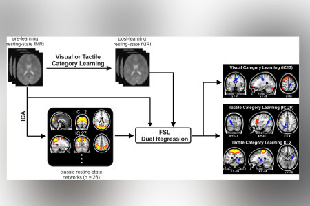

In our daily lives, we encounter new objects from various categories through different sensory modalities, such as vision and touch. Yet, how learning new categories reshapes brain connectivity networks to adapt to these objects remains poorly understood. To address this question, our research team developed an innovative paradigm that combines 3D digital embryos (artificial objects) with virtual reality and 3D-printed replicas of the same objects, simulating real-life interactions in an experimental setup.
Using dual regression analysis on resting-state fMRI data, we identified the frontoparietal network (FPN) as a shared hub modulated across both visual and tactile sensory modalities. These findings provide strong evidence for the flexible hub theory of the FPN, which suggests this network enables humans to adaptively perform a wide range of tasks by dynamically reorganizing itself.
Our study reveals that learning new categories induces sensory-modality-dependent reorganization within the brain's resting-state networks. These insights enhance our understanding of neural adaptability and may inform strategies to optimize learning through sensory experiences.


Wir gratulieren unserer Kollegin Dr. Marlies Pinnow und ihrer Gruppe herzlich zur Preisverleihung des Awards #HREA!
#HREA ist der renommierteste Award für herausragendes Personalmanagement im deutschsprachigen Raum. Am 29. November 2024 wurden in Berlin Bestleistungen in über 30 Kategorien gefeiert – und die Preisträger:innen auf großer Bühne ausgezeichnet. MAN, Barmer, Impart und die Gruppe um Dr. Marlies Pinnow wurden zusammen in der Kategorie „Wellbeing und Betriebliches Gesundheitsmanagement (BGM)" mit dem sogenannten „Flammenpokal" ausgezeichnet – und zwar mit dem Projekt Frauengesundheit er#leben in der Schwerindustrie.
Das Projekt bestand in einem mehrmonatigen betrieblichen Gesundheitsförderungs-Programm für insgesamt 50 Frauen in einem stark männerdominierten Unternehmen, das insbesondere durch beständiges Monitoring verschiedener relevanter Vitalparameter im Alltag der teilnehmenden Frauen eine Datenerhebung zum Ziel hatte. Die Auswertung dieser Daten setzt neue Maßstäbe im Bereich geschlechterspezifischer Gesundheitsförderung. Das Projekt wurde begleitet und konzipiert durch das renommierte Institute for Motivation and Personality Development Assessment Research and Training (IMPART GmbH).
Die wissenschaftliche Prä- und Postevaluation durch Mesana und die AG Motivation, geleitet von Dr. Marlies Pinnow an der Ruhr-Universität Bochum, zeigt bedeutsame positive Effekte der Maßnahmen in der Entwicklung der Selbststeuerungskompetenz der Teilnehmerinnen."
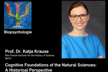

When: December 13th, 2024, at 14 c.t. | Where: IB 4-115 | Zoom: Meeting-ID 666 1535 7896, Password 390776.
Throughout history, conceptions of human cognition have significantly shaped contemporaneous understandings of the natural sciences. Questions such as what the natural sciences aim at, how they are practiced, and what their purposes are have often been answered in light of the images that the cognitive sciences—broadly construed—havecast about our human capabilities and limitations of perception, imagination, and reason. In this talk, I argue that scientists as influential as Aristotle (384-322 BCE), Albert the Great(1200-1280 CE), and Hermann von Helmholtz (1821-1894) shared and practiced this seemingly perennial insight; and I ask whether molding the specific images of human cognition in the cognitive sciences deliberately could co-shape contemporary scientific paradigms.
Short Bio: Katja Krause, professor of the history of science at the Technical University Berlin, leads the Max Planck Research Group “Experience in the Premodern Sciences of Soul and Body” at the Max Planck Institute for the History ofScience. Her research rethinks the relationship between experience and science in the premodern sciences of living beings. She is also interested in the continuities and discontinuities of scientific practices and ideals from premodernity to the present. Among her recent publications are Aquinas on Seeing God (Marquette University Press, 2020) and the edited collections Premodern Experience of the Natural World in Translation (Routledge, 2023) and Contextualizing Premodern Philosophy Explorations of the Greek, Hebrew, Arabic, and Latin Traditions (Routledge 2024). After earning her PhD in philosophy at King’s College London, Katja Krause held postdoctoral fellowships in the history of science at the MPIWG and Harvard University and an assistant professorship in medieval thought at Durham University.
View the PDF file.
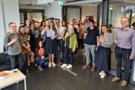

On Thursday, May 16., Alina Steinemer successfully defended her PhD thesis, entitled "Investigating avian navigation: Magnetic sense, motor projections, and the role of nitric oxide in memory formation." In her thesis, Alina examined key aspects of bird navigation using pigeons as a model species known for their exceptional homing abilities. First, Alinas showed that pigeons cannot be conditioned to respond to magnetic stimuli, nor do their brains exhibit activity in hypothesized magnetically sensitive regions. This casts a dark shadow on pigeon magnetoperception. Alina then mapped neural pathways from the nidopallium caudolaterale, revealing highly organized motor projections vital for integrating navigational information. Finally, she researched the neurochemical underpinnings of avian navigation, by mapping the distinct distribution of nitric oxide-producing neurons, as a foundation for future research on its potential role in spatial learning and navigation.
Alina's defense was a full success, and the committee (consisting of Onur Güntürkün, Jonas Rose, Martina Manns und Nikolai Axmacher) was thoroughly impressed, awarding Alinas great work with magna cum laude. Congratulations, Alina! We all are very proud of you. And we are happy, that your next journey does not take you so far away from here, but just to the lab of Nikolai Axmacher.
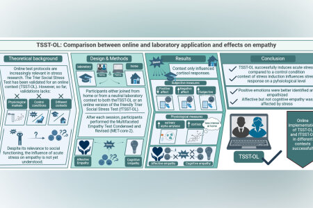

Our recent study published in Psychoneuroendocrinology examined how acute stress influences empathy using a new online stress test, the TSST-OL. This remote version of the Trier Social Stress Test was administered to 120 participants in either a lab or home setting to measure their physiological and psychological responses to stress. Researchers tracked stress markers, including cortisol and salivary alpha-amylase, along with participants' self-reported stress levels.
The findings show that while the TSST-OL reliably induced stress in both environments, cortisol responses were higher in the lab, possibly reflecting added stress from being in a formal setting. Following stress induction, participants generally had more difficulty identifying and empathizing with positive emotions, though their response to negative emotions remained unaffected. These results suggest that the home-based version of the TSST-OL might reduce anticipatory stress, making it a practical option for studying stress responses outside the lab. By validating the TSST-OL, this study highlights the potential for using remote stress testing in future research to examine how stress affects social and emotional processing.
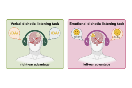

How stress affects functional hemispheric asymmetries is relevant because stress represents a risk factor for the development of mental disorders and various mental disorders are associated with atypical lateralization. Using three lateralization tasks, we investigated whether functional hemispheric asymmetries in the form of hemispheric dominance for language (verbal dichotic listening task), emotion processing (emotional dichotic listening task), and visuo-spatial attention (line bisection task) were affected by acute stress in healthy adults. One hundred twenty right-handed men and women performed these lateralization tasks in randomized order after exposure to a mild online stressor (i.e., an online variant of the Trier Social Stress Test (TSST), TSST-OL) and a non-stressful online control task (friendly TSST-OL, fTSST-OL) in a within-subjects design. Importantly, the verbal and the emotional dichotic listening tasks were presented online whereas the line bisection task was completed in paper–pencil form. During these tasks, we found the expected hemispheric asymmetries, indicating that online versions of both the verbal and the emotional dichotic listening task can be used to measure functional hemispheric asymmetries in language and emotion processing remotely. Even though subjective and physiological markers confirmed the success of the online stress manipulation, replicating previous studies, we found no stress-induced effect on functional hemispheric asymmetries. Thus, in healthy participants, functional hemispheric asymmetries do not seem to change flexibly in response to acute stress.
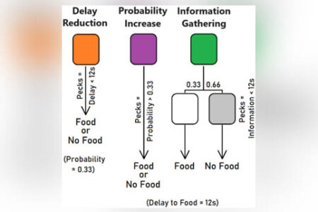

Making decisions and investing effort to obtain rewards may depend on various factors, such as the delay to reward, the probability of its occurrence, and the information that can be collected about it. As predicted by various theories, pigeons and other animals indeed mind these factors when deciding. We now implemented a task in which pigeons were allowed to choose among three options and to peck at the chosen key to improve the conditions of reward delivery. Pecking more at a first color reduced the 12-s delay before food was delivered with a 33.3% chance, pecking more at a second color increased the initial 33.3% chance of food delivery but did not reduce the 12-s delay, and pecking more at a third color reduced the delay before information was provided whether the trial will be rewarded with a 33.3% chance after 12 s. Pigeons’ preference (delay vs. probability, delay vs. information, and probability vs. information), as well as their pecking effort for the chosen option, were analyzed. Our results indicate that hungry pigeons preferred to peck for delay reduction but did not work more for that option than for probability increase, which was the most profitable alternative and did not induce more pecking effort. In this task, information was the least preferred and induced the lowest level of effort. Refed pigeons showed no preference for any option but did not drastically reduce the average amounts of effort invested. These results are discussed in the context of species-specific ecological conditions that could constrain current foraging theories.
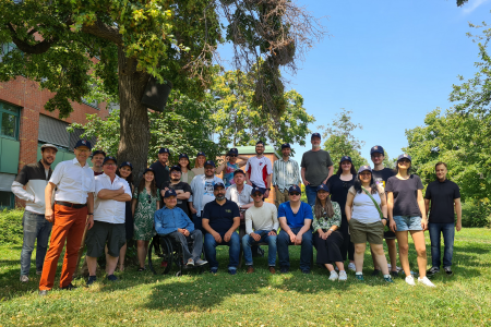

Presentation of the Future Work, SWOT by Ludwig Huber and Mike Colombo and a visit of the clever dog lab
Onur gives the plenary talk, "The unexpected evolution of neural fundaments of complex cognition"
Biopsychology lab poster session
Talk of Thomas Bugnyar and Barbara Klump, followes by a lab visit
Biopsychology Research Data Management poster and end of FENS. Jointly watching the EM quarter finale at the Donau!
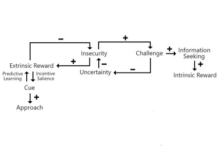

The distinction between extrinsic and intrinsic rewards and their related motivations (exploitation and exploration, respectively) has been a major concern in educational psychology for decades. Although both types of rewards are related to the dopamine-fuelled activation of the reward circuitry, neuroscientific studies now support the view that their processing also involves independent brain mechanisms. We show that these mechanisms also are already present in birds and nonhuman mammals, as they track cues and extrinsic rewards in their environment (such as food and shelter), and we discuss a number of intrinsically rewarded activities. The two categories of motivated behaviors evolved to perform distinct functions and are both crucial for the species survival. We assume that a human-animal comparison is appropriate, and suggest that both extrinsic and intrinsic rewards in humans are necessary to acquire competence, and optimally manage real-life settings, including school environments. More specifically, we argue that intrinsic and extrinsic motivations are additive rather than conflicting processes, and that intrinsic motivation is characterized by exploratory behavior and is associated with benefits for an individual; it is a step to apprehend and exploit the knowledge acquired by means of extrinsic sources of reward.
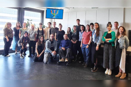

Gesa Berretz was a cornerstone of the lab and an iconic image with her colorful hairs, her Halloween outfits, and her witty charm. But at the same time, Gesa is an incredibly successful young scientist with close to 20 papers in top international journals that collect heaps of citations. Now she received a DFG grant to join the lab of Bernhard Englitz as a postdoc at Donders (Radboud University, Netherlands). But Gesa’s leave has a second implication: This now is the official end of the human group that started in 1993 and was outstandingly successful. Jointly, we changed the academic view on the mechanisms of brain asymmetries and, in close cooperation with the pigeon group, shaped a biopsychological perception on lateralization that is truly evolutionary and mechanistic. Gesa, we will miss you! All the very best.
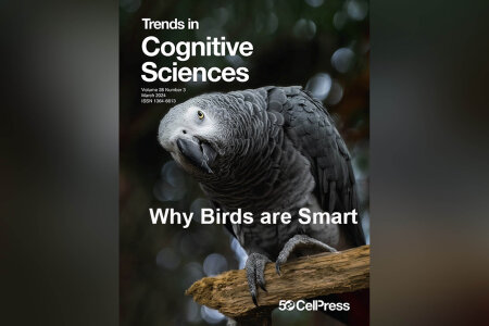

Many cognitive neuroscientists believe that both a large brain and an isocortex are crucial for complex cognition. Yet corvids and parrots possess non-cortical brains of just 1–25 g, and these birds exhibit cognitive abilities comparable with chimpanzees, which have brains of about 400 g. This paper offers an explanation for this riddle and proposes four features that may be required for complex cognition. These four neural features have convergently evolved in birds and mammals and may therefore represent ‘hard to replace’ mechanisms enabling complex cognition.
Güntürkün, O., Pusch, R., Rose, J., Why birds are smart, Trends Cogn. Sci., 2024, 28: 197-209.
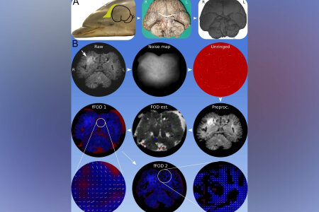

Invasive neuronal tract tracing is prohibited in very large or endangered animals, particularly marine mammals like dolphins. Scientists from Germany and Italy propose diffusion-weighted imaging as a feasible alternative, even in brains fixed in formalin for extended periods. The results of employing the Constrained Spherical Deconvolution algorithm on diffusion data from fixed bottlenose dolphin brains, demonstrated a successful identification of fiber patterns within voxels, shedding light on cetacean neuroanatomy. These findings underscore the importance of short-term post-mortem fixation for preserving tissue integrity and highlight the significance of preprocessing techniques in mitigating imaging artifacts, ultimately enhancing our understanding of cetacean brain structure.
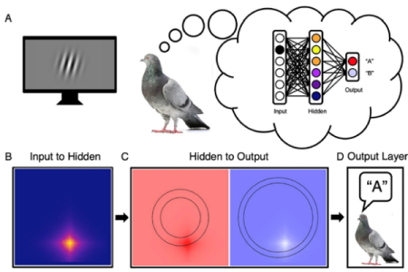

Pigeons can categorize innumerable naturalistic and artificial visual stimuli they encounter. Computational models suggest that elementary associative learning lies at the root of the pigeon’s category learning and generalization. In addition, ongoing computational and neuroscientific investigations suggest how naturalistic and artificial stimuli may be processed along the pigeon’s visual pathway. Given the pigeon’s availability and affordability, there are compelling reasons for this animal model to gain increasing prominence in contemporary neuroscientific research.
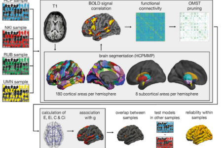

How is intelligence related to the dynamic interaction of billions of neurons in the human brain? A prominent hypothesis posits that general intelligence is related to the efficiency of the brain’s global functional network. But some publications question this link. Now, biological psychologists from Germany and the USA examined associations between g and the brain’s functional network properties in four independent data sets, by using a multi-center approach that comprises more than 2000 healthy participants. The results were sobering: No robust relation between general intelligence and the efficiency of the brain’s intrinsic functional architecture could be discovered. What is the solution? It is not clear yet, but it could be that functional connectivity is indeed relevant for intelligent thinking but only when it is investigated in a dynamic manner during conducting tasks.
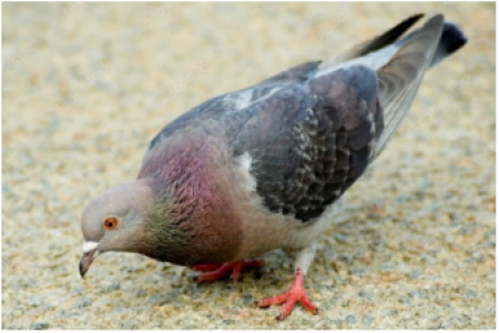

When well-known food resources are running out, animals extinguish their foraging behavior in that food patch and increasingly work for reward-related information. This re-orientation serves to decrease outcome uncertainty of foraging. To this end, animals start spending more time and effort searching for other profitable locations. This phenomenon is well known from extinction learning experiments conducted in conventional conditioning chambers where extinction ignites all kinds of novel behaviors. Now Bochum biopsychologists could show that such predictions of extinction learning research also accord with ecological observations when tested under semi-natural foraging situations. This opens new avenues of testing extinction learning mechanisms as components of ecological behavior.
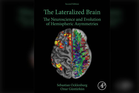

In 2017, Sebastian Ocklenburg & Onur Güntürkün published their textbook “The Lateralized Brain: The Neuroscience and Evolution of Hemispheric Asymmetries”.
While the book is an established teaching and research resource on hemispheric asymmetries by now, science does never stand still and many exciting studies on hemispheric asymmetries have been published since 2017.
Therefore, Sebastian and Onur completely overhauled their book and included hundreds of new references, many new pictures and a completely new chapter on asymmetries and development.
Like the first edition, the second edition of the book has a strong focus on integrating research findings across fields and species to give a view on hemispheric asymmetries that is informed by both neurobiology and evolution.
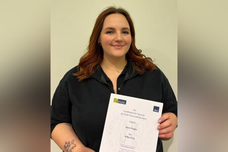

On February 2, 2024, Marie Ziegler was honored with the Wilke Award as the top graduate of her cohort in the Master’s program in Biochemistry. Presented by the Society of Friends of Ruhr University Bochum, this award recognizes Marie's exceptional academic achievements and her significant contributions to the field.
Marie’s master’s thesis, titled "Neuronal Signature of Spatial Memory in the Hippocampal Formation of Homing Pigeons", further exemplifies her dedication to advancing research in biochemistry and neurobiology.
Marie, the entire Biopsychology lab and the Faculty of Psychology at RUB are incredibly proud of you!


It is a venerable tradition that each faculty of the Ruhr University selects one of its students for the science award of the year. This usually takes place in the Auditorium Maximum as part of a large year’s end celebration. On November 29, 2023, this award was given to Robert Willma. In his master thesis, Robert had meticulously described that pigeons use a pecking code during the delay periods of working memory tasks to not forget the colors that they saw. Thus, the animals use a symbolic motor code for a percept!
Robert, the whole biopsychology lab and the faculty of psychology of the RUB is proud of you!


On Friday, December 15, 2023, Doro successfully defended her PhD thesis entitled "The neurogenetic basis of human intelligence - how network connectivity mediates the effect of polygenic scores on intelligence". Yes, it’s a long title. But it’s also a very complex topic that requires care, hard work and (of course) intelligence. In short, Doro aimed to understand how genes influence intelligence via altering the brain. She discovered that this seems to work mostly by increasing cortical surface area and the efficiency of intracortical wiring. These and many more insights prompted her to formulate a preliminary vision of a new theory for the genetic and neural fundaments of human intelligence. What an achievement!!! The committee members (Dirk Scheele, Sebastian Ocklenburg, Onur Güntürkün, Robert Kumsta) were totally impressed and awarded Doro with a summa cum laude.
Congratulations Doro, we all are very proud of you!!
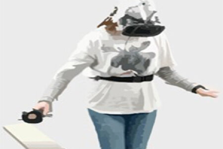

It is said that pirates made their victims walk the plank over the side of their ship. Biopsychologists from Bochum, Amsterdam, Hamburg, and Wellington made the same for science. Well, sort of. The aim was to study the neural fundaments of fear. This is usually done by showing pictures with highly negative content, but the effects are often shallow. To improve ecological validity, the scientists now asked the participants to wear virtual reality (VR) systems while walking a real plank. Looking down, they saw that the plank went over the side of an 80 storeys skyscraper. That induced true fear. And indeed, walking the plank induced a higher right hemispheric activation, as expected for fear, than looking at negative pictures. Thus, testing participants under more ecological conditions can uncover mechanisms of the emotional brain.


Some reports argued that birth complications can increase the occurrence of left handedness and thereby the risk for mental disorders. Is that true? Scientists from Bochum, Hamburg, and Göttingen joined forces and tested 598 healthy participants in depth for functional asymmetries, birth events, and clinical conditions. It turned out that early life factors had no substantial effects on functional asymmetries of hand, foot, and language. Also, the link to clinical conditions was weak. If at all, an overall reduction or an absence of asymmetries, as in mixed-handed people, seemed to be of (limited) clinical relevance.
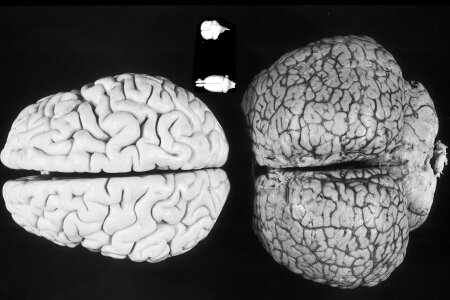

Dolphins are famous for their remarkable cognitive abilities. So, we would expect them to have a large prefrontal cortex (PFC). But there seems to be none! At least at the expected place in the frontal cortex. How is that possible? Now biopsychologists and clinical scientists from Bochum as well as veterinary scientists from the university of Padua could solve the mystery. Using MRI, they tracked the fibers ascending from the medio-dorsal thalamic nuclei (MD) to the cortex. In land mammals the MD projects to the PFC. To their surprise, they discovered that these fibers ascended to the lateral frontal cortex. So, possibly dolphins have a large PFC, but since their large brain could not grow even more frontally, their PFC was pushed sideways during evolution. The picture shows a dolphin brain along with the brains of a rat, a pigeon and the model of a human brain.
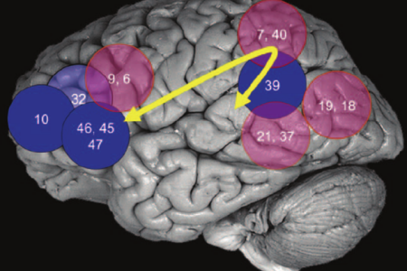

What are the neural fundaments of intelligence? According to a prominent theory, a tight interaction between some frontal and parietal areas (as shown in the picture below) could constitute these fundaments. Scientists from Bochum, Dortmund, Minneapolis, Mannheim, Edinburgh, and Minneapolis now tested a core assumption of this theory, namely that more efficient axonal pathways between these areas should go along with higher intelligence. In a multicenter approach with over 2000 healthy participants, they indeed could show that high intelligence correlates with efficiency of three critical pathways that connect these areas and their frontal interhemispheric link.
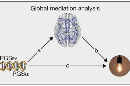

Intelligence is highly heritable. But you need to know the identity of thousands of alleles to modestly predict intelligence. Polygenic scores (PGS), which combine these effects into one genetic summary measure, are therefore used to investigate polygenic effects in intelligence research. But how do genes make brains smart? Scientists from Dortmund, Bochum, Berlin, Hamburg, Mannheim, and Luxembourg could show that intelligent individuals had larger cortical surface areas and more efficient cortical fiber connectivities, especially in parieto-frontal regions. These findings are a crucial step forward in decoding the neurogenetic underpinnings of intelligence.


Research should move into the field, into real life. But this creates a lot of problems. For example, studies related to stress have to obtain saliva samples. But swift freezing of these samples is not always possible, and samples must be returned to the laboratory for further processing. Therefore, scientists from Bochum and Hamburg tested their samples with multiple cycles of freezing and thawing, under bitter cold and scorching heat conditions, lengths of days of transport, or settings of postal delivery. By this, they developed a guideline of boundary conditions within which samples stay stable. So, now it’s time to move to the field for study!
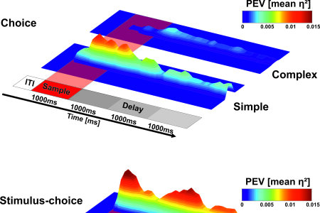

In a series of behavioral experiments complemented by single-unit recordings in pigeons (Columba livia), we show how visual information affects working memory performance. Behaviorally, complex pictures were associated with higher working memory performance compared to uniform gray pictures. This effect was accompanied by a high-dimensional multiplexed neuronal code for the representation of complex stimuli. When processing gray stimuli, neurons exclusively represented the upcoming choice. The prolonged and low-dimensional representation of the gray pictures resulted in a decay of the memory trace and a decrease in performance. In contrast, high stimulus complexity is associated with persistent neuronal multiplexing, resulting in enhanced working memory performance.
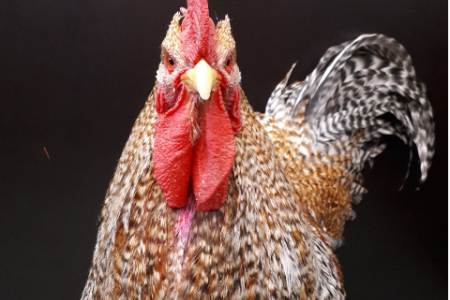

Touching a mark on the own body when seeing this mark in a mirror is regarded as a correlate of self-awareness and seems confined to great apes and a few further species. However, this paradigm often produces false-negative results and possibly dichotomizes a gradual evolutionary transition of self-recognition. We hypothesized that this ability is more widespread if ecologically tested and developed such a procedure for a most unlikely candidate: chickens (Gallus gallus domesticus). Roosters warn conspecifics when seeing an aerial predator, but not when alone. Exploiting this natural behavior, we tested individual roosters alone, with another male, or with a mirror while a hawk’s silhouette flew above them. Roosters mainly emitted alarm calls in the presence of another individual but not when alone or seeing themselves in the mirror. In contrast, our birds failed the classic mirror test. Thus, chickens possibly recognize their reflection as their own, strikingly showing how much cognition is ecologically embedded.
Here you can find a report by the New York Times.


Vergessen, Erinnern, Verlernen, neu Lernen: Es braucht die ganze Vielfalt der Psychologie und der Neurowissenschaft, um den Rätseln dieser Prozesse auf die Spur zu kommen. Einen Abend lang soll es genau um diese Prozesse gehen, bei den Brain Talks im Schauspielhaus Bochum am 7. Oktober 2023 um 19.30 Uhr.
An diesem Abend verlassen junge Forschende des Sonderforschungsbereichs (SFB) 1280 Extinktionslernen ihre Labore und begeben sich auf das Sofa auf der Bühne zu Prof. Dr. Onur Güntürkün, Leiter der Abteilung Biopsychologie an der Ruhr-Universität. Unbedingt unterhaltsam, spannend interdisziplinär und prickelnd wie nur Wissenschaft es kann, stellen sie neueste Forschung und ihre Faszination daran vor.
Tickets gibt es ab sofort beim Schauspielhaus Bochum für 15 Euro, ermäßigt 8 Euro.
Sie können auch das Infoposter und die Veranstaltung auf Facebook einsehen.


Der Doktorand begeistert sich für den Zusammenhang zwischen Körper und Gehirn, zwischen Verhalten und Denken.
Was ist für Sie das Faszinierendste an der Neurowissenschaft?
Mich begeistert der Zusammenhang zwischen Körper und Gehirn, zwischen Verhalten und Denken und wie sich wesentliche kognitive Prozesse zwischen Menschen, aber auch zwischen uns und anderen Tieren unterscheiden. Wenn ich beispielsweise Einkaufen gehe und dabei meine Mahlzeiten für die kommende Woche plane, treffe ich zahlreiche komplexe Entscheidungen und Abwägungen. Vielleicht denke ich gerade an den nächsten Urlaub und wie ich bis dahin die Figur halte oder ich will jemandem eine Freude bereiten und muss den Einkauf auf die Vorlieben anderer abstimmen.
Es ist allerdings ein Fehler, davon auszugehen, dass andere Menschen und andere Tiere, wenn sie ähnliches Verhalten zeigen wie ich, dabei auch ähnliche Denkprozesse haben müssen. In der Neurowissenschaft sind diese Unterschiede sehr schön messbar, indem man nicht nur die Lösungen miteinander vergleicht, sondern auch die Rechenwege dahin, wie in der Schule damals auch. Denkprozesse sind nicht auf das Gehirn beschränkt.
Was war bislang Ihr aufregendstes Forschungsergebnis?
In den vergangenen Jahren habe ich versucht, das Verhalten von Tauben möglichst genau zu messen, hauptsächlich mit Machine Learning, Videoanalyse und 3D-Rekonstruktionen der Körperbewegungen von Tieren. Mit diesen neuen Methoden versuchen wir, ein höheres Level an Messgenauigkeit zu erreichen, machen aber auch neues Verhalten messbar, das den Denkprozess (oder den Rechenweg) abbildet und nicht nur die Lösung oder die Entscheidung.
Wir sehen in unseren Versuchen, dass Tauben abwechslungsreicheres Verhalten zeigen als das, was für das Lösen bestimmter Aufgaben notwendig und zu erwarten ist. Zum Beispiel untersucht eine Taube entfernte Futterstellen mit ausgeprägten Kopfbewegungen, die auf visuelle Aufmerksamkeit hindeuten, entscheidet sich aber letztendlich dagegen und bleibt an der nähergelegenen Alternative. Solche unterdrückten Entscheidungen konnten mit üblichen Messmethoden nicht berücksichtigt werden, weil sich die Taube nicht im Raum bewegt, oder nicht physisch mit der alternativen Futterstelle interagiert.
Ein weiteres Beispiel ist, wenn Tauben ein individuelles und vor allem aufgabenunabhängiges Verhaltensmuster entwickeln, um bestimmte Informationen im Gedächtnis zu behalten. Sie können ihren Körper in eine bestimmte Richtung drehen, den Kopf auf eine bestimmte Weise neigen oder an einer bestimmten Stelle picken und das mit unterschiedlicher Frequenz und Stärke, um eine Entscheidung zu kodieren. Vielleicht ist dieses Verhalten vergleichbar mit meinen Fingerbewegungen beim Kopfrechnen.
Diese Ergebnisse müssen noch genauer untersucht werden, aber bisher passt das sehr gut zur Theorie und Forschungsrichtung der „verkörperten Kognition“ oder Embodied Cognition. Denkprozesse sind danach nicht auf das Gehirn beschränkt, sondern finden in direkter Interaktion mit dem Körper und der Umwelt statt, in Bewegung und Verhalten. Inzwischen denke ich häufiger auf Deutsch als auf Spanisch.
Sie haben an einer deutschen Schule in Barcelona Abitur gemacht. Wie kam es zu dieser Verknüpfung mit Deutschland, die Sie nacheinander nach Dortmund, Wuppertal und Bochum führte?
Damit hatte ich selbst nicht viel zu tun. Meine Eltern sprechen bis heute tatsächlich kein Deutsch, haben mich aber schon im Kindergartenalter in der Deutschen Schule in Barcelona eingeschult, vielleicht als Experiment? Ab da hat es mich konsistent nach Deutschland gezogen und in wenigen Wochen feiere ich mein zwölfjähriges Einwanderungsjubiläum. Deutschland im Allgemeinen, aber insbesondere NRW, sind ein großartiger Ort zum Leben, Studieren und Forschen. Die Arbeitsgruppen in Bochum um Professor Onur Güntürkün und Professor Albert Newen sind tatsächlich sehr international, aber inzwischen denke ich häufiger auf Deutsch als auf Spanisch.
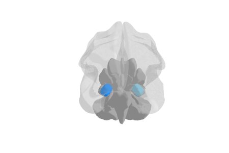

They focused on an important problem: Results of neuroimaging studies on the amygdala are often heterogeneous since it is composed of functionally and neuroanatomically distinct subnuclei. Fortunately, ultra-high-field imaging offers more accurate functional and structural representations of subnuclei and their connectivity. Most clinical studies using ultra-high-field imaging focused on major depression, suggesting either overall rightward amygdala atrophy or distinct bilateral patterns of subnuclear atrophy and hypertrophy. Connectivity analyses identified widespread networks for learning and memory, stimulus processing, cognition, and social processes, thereby provide evidence for distinct roles of different amygdala subnuclei. The researchers propose theoretical and methodological considerations that will help future ultra-high-field imaging to disentangle the ambiguity of the amygdala’s function, structure, connectivity, and clinical relevance.
Kirstein CF, Güntürkün O, Ocklenburg S. (2023). Ultra-high field imaging of the amygdala - A narrative review. Neurosci Biobehav Rev, 152, 105245.
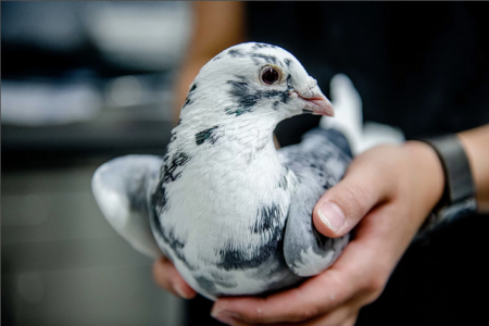

Dreaming has long been considered something that characterises human sleep. However, new findings suggest that pigeons may experience flight scenes during sleep. Mammalian sleep has been implicated in maintaining a healthy extracellular environment in the brain. During wakefulness, neuronal activity leads to the accumulation of toxic proteins, which the glymphatic system is thought to clear by flushing cerebral spinal fluid (CSF) through the brain. In mice, this process occurs during non-rapid eye movement (NREM) sleep. In humans, ventricular CSF flow has also been shown to increase during NREM sleep, as visualized using functional magnetic resonance imaging (fMRI). The link between sleep and CSF flow has not been studied in birds before. Using fMRI of naturally sleeping pigeons, we show that REM sleep, a paradoxical state with wake-like brain activity, is accompanied by the activation of brain regions involved in processing visual information, including optic flow during flight. We further demonstrate that ventricular CSF flow increases during NREM sleep, relative to wakefulness, but drops sharply during REM sleep. Consequently, functions linked to brain activation during REM sleep might come at the expense of waste clearance during NREM sleep.


The climate crisis has a massive impact on human health and mental health. Our new essential describes how brain structure and function are influenced by changes in the environment and explains the importance of ecological niches for the development of the brain. We also describe how consequences of the climate crisis, like extreme heat waves, floods, and loss of biodiversity, influence our mental health and our brain. Furthermore, we call for an urgent need for action in health sector and science to mitigate climate change related health risks.
Metzen, D., & Ocklenburg, S. (2023). Die Psychologie und Neurowissenschaft der Klimakrise – Wie unser Gehirn auf Klimaveränderungen reagiert. Springer Berlin, Heidelberg. doi: 10.1007/978-3-662-67365-2


Träumen galt lange Zeit als etwas, das den Schlaf des Menschen auszeichnet. Neue Erkenntnisse deuten jedoch darauf hin, dass Tauben im Schlaf möglicherweise Flugszenen erleben. Wissenschaftler*innen der Ruhr-Universität Bochum und des Max-Planck-Instituts für biologische Intelligenz untersuchten mithilfe der funktionellen Kernspintomographie die Aktivierungsmuster im Gehirn schlafender Tauben. Die Studie zeigte, dass das Taubengehirn während des REM-Schlafs größtenteils sehr aktiv ist. Dieser wachähnliche Zustand könnte jedoch zur Folge haben, dass das Gehirn nur unzureichend von schädlichen Substanzen gereinigt wird. Die Studie erschien in der Zeitschrift Nature Communications am 5.6.2023.
Während wir schlafen, durchläuft unser Gehirn eine Reihe komplexer Prozesse, die dafür sorgen, dass wir erholt aufwachen. Beim Menschen gehen die unterschiedlichen Schlafphasen, der REM-Schlaf (Rapid Eye Movement) und der Non-REM-Schlaf, mit Veränderungen in der Physiologie, der Gehirnaktivität und des Bewusstseins einher. Während des REM-Schlafs ist unser Gehirn besonders aktiv und wir erleben lebhafte, bizarre oder emotionale Träume. In der Non-REM-Schlafphase ist das Gehirn metabolisch weniger aktiv und entsorgt Abfallprodukte. Dazu spült es die Gehirn-Rückenmarksflüssigkeit durch die miteinander verbundenen Hirnkammern, welche die Gehirnstrukturen umgeben, und anschließend in das Gehirn. Dieser Prozess unterstützt den Körper vermutlich dabei, schädliche Proteinablagerungen, die unter anderem an der Entstehung der Alzheimer-Erkrankung beteiligt sind, aus dem Gehirn zu spülen.
Ob ähnliche Vorgänge auch bei Vögeln ablaufen, blieb bisher ungeklärt. „Der letzte gemeinsame evolutionäre Vorfahre von Vögeln und Säugetieren lebte vor etwa 315 Millionen Jahren und stammt somit aus der Frühzeit der Landwirbeltiere“, sagt Prof. Dr. Onur Güntürkün, Leiter der Biopsychologie an der Ruhr-Universität Bochum. „Dennoch ähneln die Schlafmuster von Vögeln denen der Säugetiere auf erstaunliche Weise, und zwar sowohl in den REM- als auch in den Non-REM-Phasen.“
Um herauszufinden, was genau vor sich geht wenn Vögel schlafen, haben die Forschenden die Schlaf- und Wachzustände von 15 Tauben mit Infrarot-Videokameras und funktioneller Kernspintomographie (fMRT) beobachtet und aufgezeichnet. Die Vögel waren speziell darauf trainiert, unter diesen experimentellen Bedingungen zu schlafen.
Die Videoaufzeichnungen gaben Aufschluss über die jeweilige Schlafphase der Vögel. „Wir konnten beobachten, ob nur ein oder beide Augen im Schlaf geöffnet oder geschlossen waren. Durch die transparenten Augenlider der Budapest-Tauben konnten wir auch bei geschlossenem Lid die Augenbewegungen und Veränderungen der Pupillengröße messen“, erklärt Mehdi Behroozi aus dem Bochumer Team. Gleichzeitig lieferten die fMRT-Aufzeichnungen Informationen über die Hirnaktivierung und die Bewegung der Gehirn-Rückenmarksflüssigkeit durch die Hirnkammern.
Hier geht es zu der Meldung auf der Seite des Max-Planck-Institutes
Original-Paper: https://doi.org/10.1038/s41467-023-38669-1
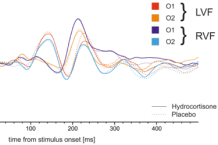

It might be presumed that due to the presence of two distinct hemispheres, each hemisphere operates autonomously without any interaction with the other. However, it is important to note that the hemispheres continually exchange information through the corpus callosum, a bundle of white matter that connects them.
The integration of information across the corpus callosum can be influenced by hormones like estradiol and progesterone. The objective of the present study was to examine whether acute stress and its accompanying fluctuations in stress hormone levels impact the transfer of information across the corpus callosum.
For this purpose, we collected EEG data from 50 participants while completing a lexical decision task and a Poffenberger paradigm twice, once after the administration of 20mg of hydrocortisone and once after a control condition. There were no differences in interhemispheric transfer between the experimental and the control in any of the tasks used to measure information exchange between the hemispheres. This suggests that increases in cortisol levels alone are not sufficient to affect interhemispheric interaction through the corpus callosum.
Berretz G, Packheiser J, Wolf OT and Ocklenburg S (2023) A single dose of hydrocortisone does not alter interhemispheric transfer of information or transcallosal integration. Front. Psychiatry 14:1054168. doi: 10.3389/fpsyt.2023.1054168


The renewed focus on avian neuroscience has sparked an explosion of new data in the field. At the same time, our understanding of sensory and particularly visual structures in the avian brain has shifted fundamentally. We review the contribution of avian categorization research—at the methodical, behavioral, and neurobiological levels. A behavioral perspective on avian categorization, is complemented by a description of the functional and structural organization of the avian visual system. Recent anatomical discoveries and the new perspective on the avian 'visual cortex' leads to the neurocomputational basis of perceptual categorization in the bird's visual system. Beyond the visual system, the "avian prefrontal cortex" and its contribution to perceptual categorization as well as insights how brain asymmetries contribute to categorization culminate in a mechanistic view of the neural principles of avian visual categorization and its putative extension to concept learning.


Birds are excellent model organisms to study perceptual categorization and concept formation. As part of the 25th Anniversary Special Issue of Animal Cognition, we review recent discoveries that have revealed how categorization is mediated in the avian brain and propose a theoretical framework that goes beyond the realm of birds. The renewed focus on avian neuroscience has sparked an explosion of new data in the field. At the same time, our understanding of sensory and particularly visual structures in the avian brain has shifted fundamentally. We review the contribution of avian categorization research—at the methodical, behavioral, and neurobiological levels. A behavioral perspective on avian categorization, is complemented by a description of the functional and structural organization of the avian visual system. Recent anatomical discoveries and the new perspective on the avian 'visual cortex' leads to the neurocomputational basis of perceptual categorization in the bird's visual system. Beyond the visual system, the "avian prefrontal cortex" and its contribution to perceptual categorization as well as insights how brain asymmetries contribute to categorization culminate in a mechanistic view of the neural principles of avian visual categorization and its putative extension to concept learning.
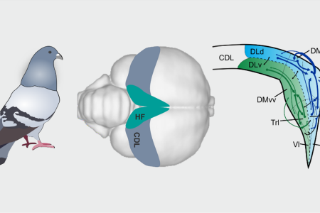

Many bird species have outstanding spatial abilities as visible during migration, homing or food caching. Just like in mammals, these spatial functions rely heavily on the integrity of the hippocampus (HC). Interestingly, recent studies reporting place cells and sharp wave ripples in birds find physiological differences between dorsomedial and ventrolateral hippocampal regions, suggesting a laminar organization along the transverse axis that seemingly stands in contrast to the non-laminar appearance of the avian HC. Here, we investigated the intrinsic connectivity of the pigeon HC especially with respect to this potential segregation of subdivisions. Using in vivo and high-resolution in vitro tracings, as well as by analyzing the neurochemical profile, we indeed identified two distinct processing streams along the avian hippocampal transverse axis that might have different functional specializations. Overall, our study suggests that the avian HC contains a complex intrinsic network suitable to sustain and modulate incoming neural activity by a recurrent neural network.

Behavior is the key to the mind, but can machines recognize and classify behavior by automated means? This is what biopsychologists and computer scientists could demonstrate by using the deep-learning-based pose-estimation tool DeepLabCut. By establishing supervised machine learning predictive classifiers for pigeon behaviors, they created an open-source software package that recognizes eating, walking, head shaking, tail shaking, preening, and fluffing with high precision and could thus analyze time-series analyses of behavioral sequences.
Wittek, N., Wittek, K., Keibel, C., Güntürkün, O., Supervised machine learning aided behavior classification of pigeons, Behavior Research Methods, 2022, https://doi.org/10.3758/s13428-022-01881-w
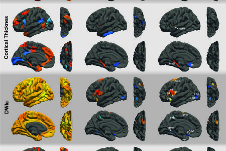

Intelligence is highly heritable. Genome-wide association studies (GWAS) have shown that thousands of alleles contribute to variation in intelligence with small effect sizes. Polygenic scores (PGS) can be used to combine these effects into one genetic summary measure. With PGS it is possible to investigate genetic predispositions in smaller, independent samples. While the genetic association of PGS with intelligence have been shown multiple times, it is largely unknown how brain structure and function are working as a neurobiological pathway behind these genetic effects. Here, we show that individuals with higher PGS for educational attainment and intelligence had higher scores on cognitive tests, larger surface area, and more efficient fiber connectivity derived by graph theory. Efficiency of fiber connectivity means that brains are organized in a way that information can easily be passed from one brain area to another with relatively energy needed. Fiber network efficiency as well as the surface of brain areas partly located in parieto-frontal regions were found to mediate the relationship between PGS and cognitive performance. Thus, genes associated with intelligence shape surface area and fiber network efficiency of our brain and these properties then in turn facilitate our intelligent thinking. These findings are a crucial step forward in decoding the neurogenetic underpinnings of intelligence, as they identify specific regional networks that link polygenic predisposition to intelligence.
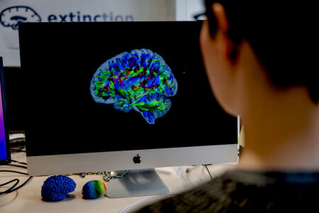

Intelligenz ist zum Teil genetisch bedingt. Es gibt Studien, die belegen, dass gewisse Genvariationen mit besseren Leistungen in Intelligenztests verknüpft sind. Andere Studien zeigen, dass unterschiedliche Hirneigenschaften, zum Beispiel eine effiziente Vernetzung, mit Intelligenz zusammenhängen. Erstmals haben Forschende nun alle drei Parameter – Gene, unterschiedliche Hirneigenschaften und Verhalten – gleichzeitig untersucht. Mit Genanalysen, kernspintomografischen Aufnahmen und Intelligenztests wies das Team nach, welche Hirneigenschaften das Bindeglied zwischen Genen und Verhalten bilden.
Die Ergebnisse beschreibt ein Team um Dorothea Metzen von der Arbeitseinheit Biopsychologie der Ruhr-Universität Bochum und Dr. Erhan Genç, früher an der Ruhr-Universität, heute am Leibniz-Institut für Arbeitsforschung in Dortmund (IfADo), in der Zeitschrift „Human Brain Mapping“, online veröffentlicht am 4. April 2023.
Neben dem IfADo und verschiedenen Einrichtungen der Ruhr-Universität waren die Humboldt-Universität Berlin, das Zentralinstitut für Seelische Gesundheit in Mannheim, die Medical School Hamburg und die Universität Luxemburg beteiligt. An der Ruhr-Universität kooperierten die Teams der Biopsychologie, der Humangenetik und der Genetischen Psychologie.
Hier geht es zu der Meldung auf der "Rub News" Seite.
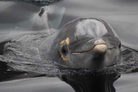

Mirror-guided self-recognition is seen as a cognitive hallmark that possibly indicates knowledge about oneself. Only a few species have up to now passed a standard mark-and-mirror test in which animals attempt to touch a mark that can only be seen in the mirror. Mirror self-recognition has also been claimed for bottlenose dolphins but this initial work is highly contested. The problem is: “How do test an animal that cannot touch the mark because it has no arms?” To solve this problem, experts of dolphin behavior in St. Andrews, Nuremberg, and Bochum invented a new procedure in which they marked the areas around both eyes of the animals at the same time, one with visible and the other with transparent dye to control for haptic cues. This allowed the animal to see the mark easily and the scientists to investigate what side the animal looked at when being confronted with a mirror. They found that the animals actively chose to inspect their visibly marked side while they did not show an increased interest in a marked conspecific in the pool. These results make it likely that dolphins can differentiate between self and others.


In humans, stress can alter functional asymmetries. But is this also true for other species like, e.g., dogs, our companion since Thousands of years? The tested dogs were either from private pet owners in the region of Ankara or working dogs from an official dog training tenter. A vet determined which animals showed or did not show signatures of chronic stress. Mild acute stress was induced by placing the animals in an open field. The results clearly indicated that acute and chronic stress reduced natural functional asymmetries in dogs. Thus, such a pattern seems to be a general mechanism and could be used to probe stress levels of animals like dogs.


On May 17, 2023, the raw construction of THINK was finalized and a very nice topping out ceremony took place. Politicians from the state of North Rhine-Westphalia and the city of Bochum as well as representatives of the building agency and our university gave short congratulation addresses. Onur shortly recapitulated the history of THINK and stressed the collaborative effort of many people who made THINK possible. THINK is 100 meters long and 45 meters wide, the floor space is 4,000m2 of which more than half is laboratory space, the four-story building is naturally lit via an inner courtyard, and the high-resolution human and small-animal magnetic resonance imaging (MRI) scanners required an unusual structural design in which the equipment foundations under the MRIs are decoupled from the foundations of the rest of the building.
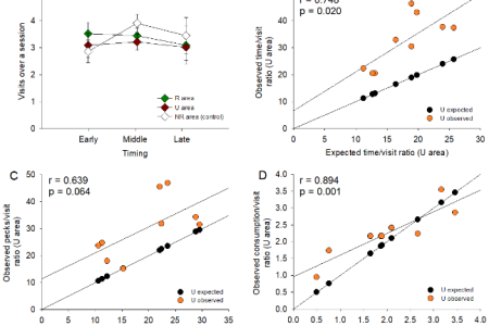

One major survival-related activity of organisms is to seek food in their environment. To this end, they exploit previously rewarding locations and attempt to approach the cues predictive of the edible items they detect or expect. But foraging is unlikely to be a matter of reinforcement only. If it was, however, foraging activity should follow principles of extinction learning: It should be abolished in a location without reinforcement and proportionally be reduced in a partially reinforced location relative to a fully reinforced one containing the same number of food items. We tested these two hypotheses using a foraging board, which allowed pigeons to find food items hidden in perforated holes. Our results showed that the overall time spent and the overall number of pecks given in one area was related to reinforcement density in that area. To a lesser extent, the same phenomenon occurred with respect to the number of visits per area. However, the time-per-visit and pecks-per-visit ratios were higher in the partially vs. fully reinforced area, suggesting that the pigeons foraged more than expected when food was uncertain. These results will be discussed in the context of the matching law and optimal foraging.
Anselme, P., Wittek, N., Oeksuez, F., Güntürkün, O. (2022). Overmatching under food uncertainty in foraging pigeons. Behavioural Processes, 201, 104728. https://doi.org/10.1016/j.beproc.2022.104728
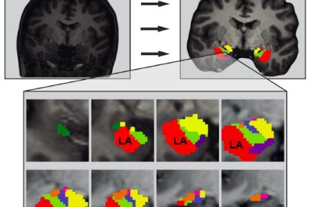

Neuroticism is a personality trait that is associated with high levels of anxiety, anger, and loneliness. Biopsychologists in Bochum have now discovered a link between the cellular microstructure of the amygdala and neuroticism. Brain images from 221 healthy participants were acquired using advanced multi-shell diffusion-weighted imaging and the amygdala was segmented into its eight subnuclei. Lower neurite density in the lateral amygdala nucleus (LA) was significantly associated with higher scores in depression, one of the six neuroticism facets. The La is the sensory relay of the amygdala, filtering incoming information based on previous experiences. Reduced neurite density and related changes in the dendritic structure of the LA could impair its filtering function, causing harmless sensory information to be misevaluated as threatening and thus increase neuroticism-related behavior.
Schlüter, C., Fraenz, C., Friedrich, P., Güntürkün, O. and Genç, E., Neurite density imaging in amygdala nuclei reveals interindividual differences in neuroticism. Human Brain Mapping, 2022, 1–13 https://doi.org/10.1002/hbm.25775.
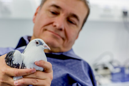

Unsere geistige Fähigkeit, die komplexe Welt in Kategorien einzuteilen, erleichtert unser tägliches Leben. Doch wie kommen wir zu dieser Einteilung? Und welche Merkmale werten wir aus? Der Beantwortung dieser Fragen sind Forschende der Ruhr-Universität Bochum mit der Hilfe von Tauben ein Stück näher gekommen. Sie fanden heraus, dass Vögel verschiedene Strategien nutzen, um Kategorien erfolgreich zu lernen. Für ihre Datenerhebung nutzen die Wissenschaftler eine neuentwickelte Forschungsmethode. Sie kombinierten dabei die sogenannte „virtuelle Phylogenese“, bei der künstliche Stimuli computergestützt erzeugt werden, mit einem maschinellen Lernansatz und somit der automatisierten Auswertung des Pickverhaltens der Vögel. Die Erkenntnisse ihrer Forschung veröffentlichten sie nun in der Januarausgabe der Zeitschrift Animal Cognition.
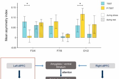

Each half of the brain is specialized for processing different tasks. The left hemisphere is more strongly involved in language processing while the right hemisphere is dominant for face perception. For example, frontal asymmetries measured with EEG have been linked to both emotional processing in healthy individuals and affective disorders like depression. Stress provides a particularly strong source of negative emotion and has also been related to the pathogenesis of affective disorders. Hence, the aim of the present study was to investigate how acute stress affects frontal EEG asymmetries. For this purpose, continuous EEG data were acquired from 51 healthy adult participants during stress induction with the Trier Social Stress Test. In addition, EEG data were also collected during a non-stressful control condition. Furthermore, EEG resting state data were acquired after both of these conditions. In the stress condition, participants showed stronger left hemispheric activation over frontal electrode sites as well as reduced left-hemispheric activation over occipital electrode sites compared to the non-stressful control condition. There were no stress-related changes in the subsequently recorded resting state data. Our results are in line with predictions of the asymmetric inhibition model, which postulates that the left prefrontal cortex inhibits negative distractors, like the negative emotions resulting from acute stress. Moreover, the results support the capability model of emotional regulation, which states that frontal asymmetries during emotional challenge are more pronounced compared to asymmetries during rest conditions. These models suggest that left frontal activity could be indicative of stronger emotion regulation, which is more pronounced during emotional challenge.
Berretz, G., Packheiser, J., Wolf, O. T., & Ocklenburg, S. (2022). Acute stress increases left hemispheric activity measured via changes in frontal alpha asymmetries. iScience, 103841. https://doi.org/10.1016/j.isci.2022.103841
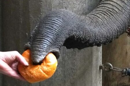

In August 2021, our PhD student John Tuff visited the lab of Prof Michael Brecht at the Bernstein Center for Computational Neuroscience Berlin, Humboldt-Universität zu Berlin as part of his Max Planck School of Cognition PhD program. In this time, he and the team from Berlin investigated the trigeminal system of the elephant brain and compared it with other sensory nerves as well as the spinal cord. They found the trigeminal ganglia to be huge, larger than a macaque monkey brain, and the maxillary branches, which lead down to the trunk, were thicker than the spinal cord, indicating that the connections to the trunk are more substantial than the nerves to the rest of the body. The infraorbital nerve, which runs through the trunk, was several times as thick as other sensory nerves. These findings suggest that while elephants are mainly known for their excellent sense of hearing, it is possibly underestimated how much these animals rely on their trunks for sensory input.
Purkart, L., Tuff, J. M., Shah, M., Kaufmann, L. V., Altringer, C., Maier, E., Schneeweiß, U., Tunckol, E., Eigen, L., Holtze, S., Fritsch, G., Hildebrandt, T. & Brecht, M. (2022). Trigeminal ganglion and sensory nerves suggest tactile specialization of elephants. Current Biology. https://doi.org/10.1016/j.cub.2021.12.051
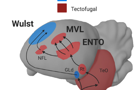

iscriminating between object categories is a crucial function of primate and bird visual systems. However, brain structures responsible for visual processing greatly differ between birds and mammals. Here, we examined whether a similar hierarchical organization that operates for processing faces in monkeys also exists in the avian visual system. While pigeons viewed images of faces, scrambled controls, and sine gratings, single neurons were recorded from three visual forebrain regions: the Wulst, the entopallium (ENTO) and mesopallium ventrolaterale (MVL). A greater proportion of single MVL neurons fired to the stimuli, and the population response of MVL neurons distinguished between stimuli with greater capacity than ENTO and Wulst neurons. In contrast to the primate system, no neurons were strongly face-selective and some responded best to the scrambled images. These findings suggest that MVL is primarily involved in processing local features of images, much like the early visual cortex.
Clark, W., Chilcott, M., Azizi, A., Pusch, R., Perry, K. & Colombo, M. (2022) Neurons in the pigeon visual network discriminate between faces, scrambled faces, and sine grating images. Sci Rep 12, 589. https://doi.org/10.1038/s41598-021-04559-z
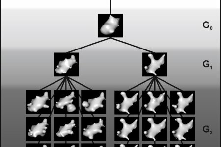

Categorizing helps us to cope with the vast variety of objects in our environment. Although categorization represents a core cognitive function, stimulus features that drive behavior and underlying strategies for categorizing objects often remain elusive. To elucidate these issues, we performed behavioral experiments with pigeons - classic model animals to investigate perceptual categorization. We generated two categories of artificial stimuli called digital embryos and analyzed the pigeons pecking behavior using machine learning. Our results show that pecking is indicative of the upcoming choice and thus related to features of interest. However, individual animals use different stimulus aspects to base their decision on. By using defined artificial stimuli in addition with a detailed analysis of the pecking behavior, our study paves the way to pinpoint stimulus features as well as individual strategies to solve the task.
Pusch, R., Packheiser, J., Koenen, C. Iovine, F. & Güntürkün, O. (2022) Digital embryos: a novel technical approach to investigate perceptual categorization in pigeons (Columba livia) using machine learning. Anim Cogn. https://doi.org/10.1007/s10071-021-01594-1
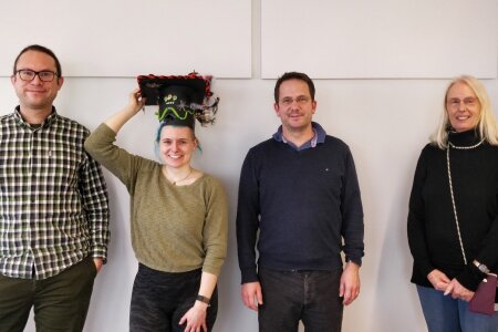

On Friday, the 17th of December 2021, Gesa successfully defended her PhD thesis entitled "How acute stress modulates hemispheric asymmetries: Investigating the role of endocrinological and affective parameters". Gesa's PhD was focused on the interplay between stress and hemispheric asymmetries and she was fittingly co-supervised by Sebastian Ocklenburg (Biopsychology) and Oliver Wolf (Cognitive Psychology). Gesa used EEG and behavioral experiments to investigate how psycho-social stress and cortisol affect hemispheric asymmetries and interhemispheric integration in healthy volunteers. Her PhD thesis included an impressive 8 manuscripts, including empirical studies, meta-analyses and a theoretical review paper that significantly advances our understanding how stress affects hemispheric asymmetries. The committee members (Oliver Wolf, Sebastian Ocklenburg, Nikolai Axmacher, Robert Kumsta) were so impressed that they awarded Gesa a summa cum laude ("with highest honors") that only very few PhD students achieve.
Congratulations Gesa! We all are very proud of you!


We all are happy that Sebastian received such a highly deserved and very well equipped position. Congratulations Sebastian! We all are very proud of you! We will immensely miss you, your famous cakes and parties, your selfless attitude of helping as well as your scientific knowledge and insights.
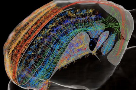

Cognitive functions are similar in birds and mammals. So, are therefore pallial cellular circuits and neuronal computations also alike? In search for answers, a group of Biopsychologists in Bochum moved in birds from pallial connectomes, to cortex-like sensory canonical circuits and connections, to forebrain micro-circuitries and finally to the avian “prefrontal” area. This voyage from macro- to microscale networks and areas reveals that both birds and mammals evolved similar neural and computational properties in either convergent or parallel manner, based upon circuitries inherited from common ancestry. Thus, these two vertebrate classes evolved separately within 315 million years highly similar pallial architectures that produce comparable cognitive functions.
Güntürkün, O., von Eugen, K., Packheiser, J., Pusch, R. Avian pallial circuits and cognition - A comparison to mammals, Current Opinion in Neurobiology, 2021, 71: 29-36. https://doi.org/10.1016/j.conb.2021.08.007
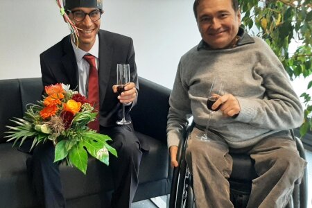

Emre conducted three PhD experiments that all resulted in nice publications. In these studies, he discovered some core principles with which the two hemispheres interact with each other through the commissura anterior under conditions of conflicting information. In a further study he solved the puzzle why pigeons can’t recognize themselves in the mirror, but can use a mirror as a tool to peek into corners that they can’t see directly. The solution for this last mystery was simple: They can’t. Instead, their nearly panoramic sight of view enables them to detect food even in areas close to their back. The committee was very happy with his achievements and was convinced that Emre fully deserves to be awarded a PhD.
Congratulations Emre! We all are very proud of you!


Neslihan’s PhD experiments are a highly innovative and technically advanced series of studies that go all the way through visual perception, action, and cognition in birds. To this end, Neslihan developed novel technical approaches to gain deep insights into diverse aspects of avian cognitive neuroscience. Two major insights stand out among many further ones. First, Neslihan discovered that the process of decision making in pigeons strongly resembles evidence accumulation curves known from mammals. Thus, this core sensory computation seems to be identical since 315 million years of separate evolution. Second, pigeons do not recognize themselves in the mirror but also don’t take their mirror image as another pigeon. Instead, for them the guy in the mirror seems to be an uncanny individual. The committee, consisting of Thomas Bugnyar, Jonas Rose, Giorgio Vallortigara, and Onur Güntürkün was highly impressed by these achievements and were convinced that she fully deserves to be awarded a PhD.
Congratulations Neslihan! We all are very proud of you!
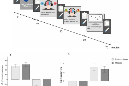

Chronic stress has been shown to have long-term effects on natural differences in function between the left and right hemisphere – so called functional hemispheric asymmetries (FHAs). The short-term effects of acute stress exposure on functional hemispheric asymmetries are less well investigated. It has been suggested that acute stress can affect FHAs by affecting the corpus callosum, the white matter pathway that connects the two hemispheres. On the molecular level, this modulation may be caused by a stress-related increase in cortisol, a major stress hormone. Therefore, a team from the biopsychology, the cognitive psychology and the department of neurology from the RUB set out to investigate the acute effects of cortisol on FHAs. Overall, 60 participants were tested after administration of 20 mg hydrocortisone or a placebo tablet in a cross-over design: participants performed a verbal and an emotional dichotic listening task to assess language and emotional lateralization respectively, as well as a Banich–Belger task to assess interhemispheric integration both times. Lateralization quotients were determined for both reaction times and correctly identified syllables in both dichotic listening tasks. In the Banich–Belger task, across-field advantages were determined to quantify interhemispheric integration. While we could replicate previously reported findings for these tasks in the placebo session, we could not detect any differences in asymmetry between hydrocortisone and placebo treatment. This partially corroborates the results of a previous study we performed using social stress to induce cortisol increases. This suggests that an increase in cortisol does not influence dichotic listening performance on a behavioral level.
Berretz, G., Packheiser, J., Höffken, O., Wolf, O. T., & Ocklenburg, S. (2021). Dichotic listening performance and interhemispheric integration after administration of hydrocortisone. Scientific Reports, 11(1), 1-11. https://doi.org/10.1038/s41598-021-00896-1
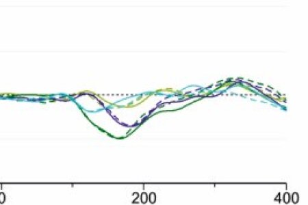

One could think that, since we have two separate hemispheres, each hemisphere works independently of the other. To a certain extent, this is true: Functional hemispheric asymmetries emerge as the left and the right hemisphere are dominant for different aspects of task processing. However, the hemispheres constantly share information through the corpus callosum, the white matter connecting between them. The integration of information across the corpus callosum is dependent on its structural integrity and functionality. Several hormones, like estradiol and progesterone, can influence this function. Since earlier work has demonstrated that long-term changes in stress hormone levels are accompanied by changes in hemispheric asymmetries in several mental disorders, the aim of the current study was to investigate whether acute stress and the associated changes in stress hormone levels also affect information transfer across the corpus callosum. For this purpose, researchers from the biopsychology and the cognitive psychology of the Ruhr University Bochum collected EEG data from 51 participants while completing a lexical decision task and a Poffenberger paradigm twice, once after stress induction with the Trier Social Stress Test and once after a control-condition. While there were no differences in interhemispheric transfer between the stress and the non-stress condition in the Poffenberger paradigm, we observed shorter latencies to stimuli in the left visual field in the left hemisphere at the CP3-CP4 electrode pair after stress. These electrodes are positioned over the brain area that is involved in lexical processing. This suggest that the transfer of lexical material from the right to the left hemisphere was quicker under stress indicative of more efficient signal transfer across the corpus callosum between language related areas.
Berretz, G., Packheiser, J., Wolf, O. T., & Ocklenburg, S. (2021). Improved interhemispheric connectivity after stress during lexical decision making. Behavioural Brain Research, 113648. https://doi.org/10.1016/j.bbr.2021.113648
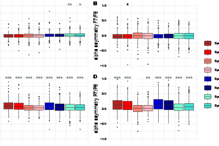

Frontal asymmetries during rest is one of the most widely investigated forms of hemispheric asymmetries. It is often assessed by measuring asymmetry in EEG resting-state alpha activation. Alpha asymmetries have been associated with important psychological outcomes like wellbeing, depression and motivation. However, studies on the reliability of alpha asymmetries are sparse and often consist of small sample sizes. In our study, we investigate short-term reliability of frontal and parietal alpha asymmetry in a large sample of 370 participants. We find that both frontal and parietal alpha asymmetry show a good reliability, especially when eyes are closed during the resting state recording. We also find, that reliability differs between recording sites, with frontomedial electrodes showing lower reliability than frontlateral and parietal sites.
Metzen, D.; Genc, E.; Getzmann, S.; Larra, M.; Wascher, E. & Ocklenburg, S. (2021). Frontal and parietal EEG alpha asymmetry: A large-scale investigation of short-term reliability on distinct EEG systems. Brain Struct Funct. https://doi.org/10.1007/s00429-021-02399-1
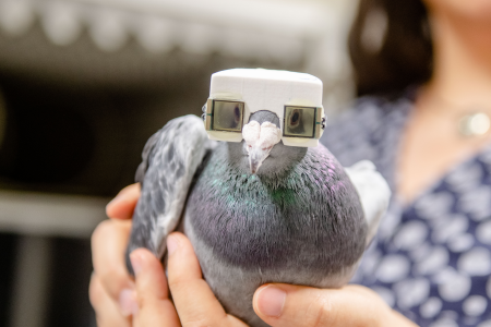

Animals constantly have to make decisions: they often have to choose between foraging strategies, mates, territories, and social partners. Thus, proper decision-making is fundamental for survival. Studies in primates and rodents revealed a stochastic perceptual evidence accumulation process during decision-making. What about birds? The present study investigated whether perceptual decision-making in pigeons shows behavioral and computational dynamics comparable to those in mammals and rodents. Using a novel "pigeon helmet" with liquid shutter displays that control visual input to individual eyes/hemispheres with precise timing, we revealed highly similar perceptual decision-making dynamics. Thus, both mammals and birds seem to share this core cognitive process that possibly represents a fundamental constituent of decision-making throughout vertebrates. We additionally discovered that avian hemispheres start independent sensory accumulation processes without any major interhemispheric exchange under conditions of time pressure.
Wittek, N., Matsui, H., Behroozi, M., Otto, T., Wittek, K., Sarı, N., Stoecker, S., Letzner, S., Choudhary, V., Peterburs, J., & Güntürkün, O. (2021). Unihemispheric evidence accumulation in pigeons. Journal of experimental psychology. Animal learning and cognition, 47(3), 303–316. https://psycnet.apa.org/doi/10.1037/xan0000290


Research into the neuronal basis of emotion processing has so far mostly taken place in the laboratory, i.e. in unrealistic conditions. Bochum-based biopsychologists have now studied couples in more natural conditions. Using electroencephalography (EEG), they recorded the brain activity of romantic couples at home while they cuddled, kissed or talked about happy memories together. The results confirmed the theory that positive emotions are mainly processed in the left half of the brain.
A group led by Dr. Julian Packheiser, Gesa Berretz, Celine Bahr, Lynn Schockenhoff and Professor Sebastian Ocklenburg from the Department of Biopsychology at Ruhr-Universität Bochum describes the results in the journal Scientific Reports, published online on 13 January 2021.
In previous studies on the neuronal correlates of emotion processing, feelings were usually triggered in subjects by presenting certain images or videos in the laboratory. “It was unclear whether that really reflects how people experience and act out feelings,” says Julian Packheiser. “Ultimately, emotions comprise not only the perception of feelings but also their expression.”
Click here to read the report on the "Neuroscience News" page.

„Der Botenstoff Dopamin ist in der Vergangenheit immer wieder mit einer erhöhten kognitiven Flexibilität in Verbindung gebracht worden“, sagt Dr. Erhan Genç aus der Bochumer Abteilung für Biopsychologie. „Das ist nicht grundsätzlich schlecht, aber geht oftmals mit einer erhöhten Ablenkbarkeit einher.“
In der Zeitschrift Social Cognitive and Affective Neuroscience, vorab online veröffentlicht am 3. Juli 2019, berichtet Erhan Genç unter anderem zusammen mit Caroline Schlüter, Dr. Marlies Pinnow, Prof. Dr. Dr. h. c. Onur Güntürkün, Prof. Dr. Christian Beste und Privatdozent Dr. Sebastian Ocklenburg über die Ergebnisse.
Die Forschungsgruppe untersuchte die genetische Ausstattung von 278 Männern und Frauen. Sie interessierten sich vor allem für das sogenannte Tyrosinhydroxylase-Gen (TH-Gen). Je nach Ausprägung des Gens besitzen Menschen im Gehirn viel oder wenig Botenstoffe aus der Katecholamin-Familie, zu denen der Botenstoff Dopamin gehört. Außerdem erfasste das Team mit einem Fragebogen, wie gut die Teilnehmerinnen und Teilnehmer ihre Handlungen kontrollieren können. Frauen mit schlechterer Handlungskontrolle hatten die genetische Anlage für höhere Dopaminlevel.
Hier geht es zum Text der Meldung auf der "Informationsdienst Wissenschaft" Seite.
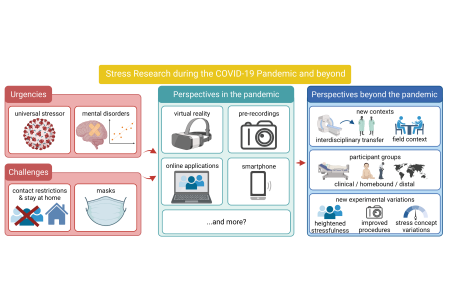

Stress research is of crucial relevance in the context of the COVID-19 pandemic. However, lockdowns and contact restrictions imposed to prevent the virus from further spreading, confront stress researchers in psychology and neuroscience with unique challenges. Experimental paradigms widely used to induce stress typically feature in-person social encounters. Hence, they conflict with COVID-19-related requirements. In order to continue stress research during the pandemic, researchers were forced to adapt established stress protocols. We reviewed the literature concerning trends and perspectives such as virtual reality, pre-recordings and online formats that may be useful to adapt established stress induction paradigms to COVID-19-related requirements. Regarding online formats, for instance, it is well feasible to transfer above-mentioned psychosocial stress induction to online communication software in where participant and investigators can meet without coming to research facilities where they may be set at risk for COVID-19 infection. Importantly, we concluded that some approaches to adapt stress protocols may not only help to continue stress research during COVID-19 but that they will likely stimulate the field far beyond the pandemic. For example, altered procedures may open stress research to new contexts or more diverse participant groups. Moreover, they bear the potential for new experimental manipulations.
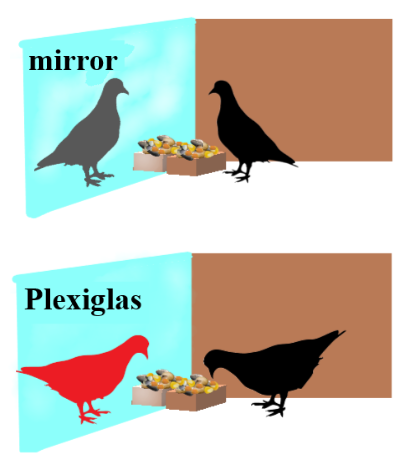

What do animals perceive when they are confronted with their mirror image? And how do answers to this question change our own understanding of consciousness and the relationship between humans and animals? While humans tend to intuitively project their own perception of the world onto other species, we set out to challenge the traditional binary model of mirror self-recognition, which assumes a sharp “cognitive Rubicon” that only a limited number of species seemingly can pass.
Earlier studies had shown that pigeons do not pass the traditional mark test, although they can detect synchronicity between self-and foreign movements. However, it was not clear whether they distinguish between self-movements in a mirror reflection and the movements of a real conspecific and behave accordingly. Thus, we tested pigeons in a non-traditional mirror self-recognition task in which they were forced to feed in front of their mirror reflection or in front of another conspecific. Detailed manual observations and automated analysis revealed that while the foreign pigeon was accepted as a competitor, the mirror image caused more inhibition as the reflection seemingly represented an uncanny individual. These results strengthened the case for a gradualist interpretation of MSR as opposed to the traditional binary model. By failing to succeed in spontaneous MSR tasks while being able to make a difference between their mirror image and unknown conspecific, pigeons belong to an intermediate category.
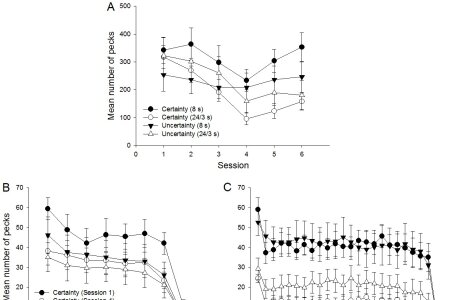

The Pavlovian autoshaping paradigm has often been used to assess the behavioral effects of reward omission on behavior. We trained pigeons to receive a food reward (unconditioned stimulus, or UCS) following illumination of a response key (conditioned stimulus, or CS). In Experiment 1, 1 group of pigeons was trained with two 100% predictive CS-UCS associations (reward certainty) and another group with two 25% predictive CS-UCS associations (reward uncertainty) for 12 sessions. In both groups, the 2 CS durations were 8 s. Then, in each group, the duration of 1 CS remained unchanged and that of the other CS was suddenly extended from 8 to 24 s for 6 sessions. In Experiment 2, some experienced individuals (from Experiment 1) and naïve individuals formed 2 groups trained with a 24-s CS throughout for 18 sessions. Our results show that pigeons (a) pecked less at the uncertain than the certain CS, (b) decreased and then increased CS-pecking after extending the CS duration, especially in the certainty condition, (c) were unresponsive to the 24-s CS in the absence of previous experience, and (d) decreased their response rate close to the end of a trial irrespective of the reinforcement condition, CS duration, and amount of training. These results are discussed in relation to several theoretical frameworks.
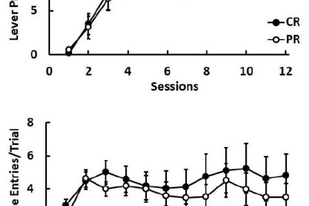

In Pavlovian autoshaping, sign-tracking responses (lever pressing) to a conditioned stimulus (CS) are usually invigorated under partial reinforcement (PR) compared to continuous reinforcement (CR). This effect, called the PR acquisition effect (PRAE), can be interpreted in terms of increased incentive hope or frustration-induced drive derived from PR training. Incentive hope and frustration have been related to dopaminergic and GABAergic activity, respectively. We examined the within-trial dynamics of sign and goal tracking in rats exposed to 20-s-long lever presentations during autoshaping acquisition under PR vs. CR conditions under the effects of drugs tapping on dopamine and GABA activity. There was no evidence of the PRAE in these results, both groups showing high, stable sign-tracking response rates. However, the pharmacological treatments affected behavior as revealed in within-trial changes. The dopamine D2 receptor agonist pramipexole (0.4 mg/kg) suppressed lever pressing and magazine entries relative to saline controls in a within-subject design, but only in PR animals. The allosteric benzodiazepine chlordiazepoxide (5 mg/kg) failed to affect either sign or goal tracking in either CR or PR animals. These results emphasize the roles of dopamine and GABA receptors in autoshaping performance, but remain inconclusive with respect to incentive hope and frustration theories. Some aspects of within-trial changes in sign and goal tracking are consistent with a mixture of reward timing and response competition.
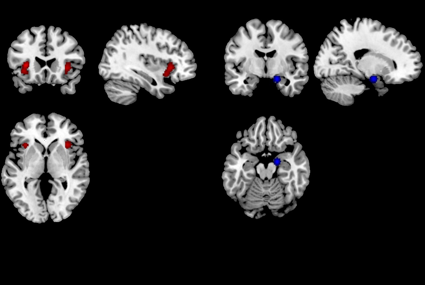

Everyone feels stress - be it a lecture at university, an interview or a first date. While we know that stress affects the entire information processing path, for example attention, working and long-term memory, there is so far no consensus on how these different situations trigger the same feeling of stress and what happens in the brain.
Many scientists try to answer this question and use different methods to induce stress in their subjects. During this, the participants' brain activity is measured. Given the variety of methods used, the question arises to what extent they are comparable with each other and which brain structures are activated by stress.
In order to answer this question, we have carried out a systematic summary and analysis of the previously published literature on the subject: In the Activation-Likelihood Estimation Analysis, ALE for short, the coordinates of the activation patterns from all studies are compared during the stress and it is statistically checked how similar the different patterns are. As a result, we get a number of areas of the brain that have shown activation in all studies.
We found cross-method activation in the insula, the claustrum, the lentiform nucleus and the inferior frontal gyrus, among others.
The insula is related, among other things, to pain perception, self-perception and social perception. As part of the so-called salience network, it integrates sensory and internal emotional information. In addition, it helps to control the hormonal stress response. The Claustrum is located directly adjacent to the insula. Similar to its neighbor, the Claustrum is also responsible for the integration of various information. It also has a meaning for consciousness. This suggests that under stress, study participants turn their attention inwardly to their emotional processes.
The inferior frontal gyrus is responsible for semantic and phonological processing as well as working memory. Since many of the procedures involve strenuous mental tasks, his role here is likely to be the processing of these tasks.
Overall, our study shows that most of the methods used to induce stress work well and produce similar results. It also shows that the insula, claustrum and inferior frontal gyrus play a central role in the stress response.
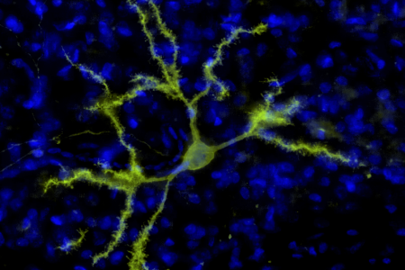

Birds are excellent models to study learning, complex cognition, song and vision. However, to understand how these behaviors are realized within the brain, methods that are able to control cellular activity on a millisecond timescale are essential. This can be achieved with optogenetics, where neurons become light-sensitive through the integration of artificial ion channels and can later be controlled with focused optical stimulation. However, this method relies on the use of viral vectors that have so far never been used in the pigeon brain. Therefore, in our study, we compared the efficiency of three adeno-associated viral vectors and found that AAV1 was the most efficient construct in delivering ChR2 into the primary visual area and various other brain regions of pigeons. We could also verify the light responsiveness of ChR2 expressing cells with electrophysiological recordings and immediate early gene expression. Most importantly, we provided the first proof of principle for optogenetics in behaving pigeons as optical stimulation of the primary visual area decreased pigeons’ grayscale visual discrimination accuracy. With our study we provide a viral strategy that can be used for the functional analysis of brain circuits in pigeons and offers novel opportunities for comparative research.

On Thursday, the 26th of November 2020, Sebastian was elected to the ranks of a professor – a title that is long overdue given his outstanding scientific achievements. Technically speaking, he assumed the title of an “Außerplanmäßiger Professor”. This requires the fulfillment of lots of academic criteria of excellence. In addition, two leading external scientists had to verify that Sebastian could successfully receive a call from their institutions. Their response letters were sparked with words like “a most creative and brilliant experimental scientist”, “at the forefront of research”, “tripled the expectation”, etc. So, it was absolutely clear that Sebastian deserves this title and the committee as well as the rectorate did not hesitate a second.
Congratulations Professor Sebastian Ocklenburg! We all are excited!


On Friday, the 30th of October 2020, Noemi successfully defended her doctoral thesis on the “Neural Circuits of Multi-Component Behavior in the Avian Brain”. Noemi delivered a fantastic tour through her research by first explaining the omnipresence of what we loosely call “multitasking” in our everyday life. She explained that different kinds of functional organizations of action selection and execution are present under this umbrella term and can be beautifully teased apart by the Stop-Change paradigm. Using this procedure, parallel and serial actions can be disambiguated. However, a major problem remains and blocks the path for causal studies: There is no animal model. The establishment of such an animal model is exactly the aim of Noemi’s thesis. To this end, she established all necessary components to show that pigeons can be perfectly tested as an animal model and can help to tease apart the critical neural components by using molecular imaging and behavioral local pharmacology. As beautiful as all of these achievements are, the crown jewel of her thesis is the subsequent establishment of optogenetics in pigeons. For sure, this achievement will change the landscape. The committee was deeply impressed and awarded this grand work with a magna cum laude.
Congratulations Noemi! We are incredibly proud of you.
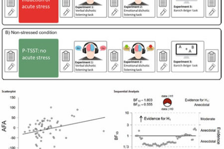

Left – analytical, right – creative: that is a misconception many people still have about the two of the halves of brain. Although this is exaggerated, both hemispheres display differences in task processing – so called functional hemispheric asymmetries (FHAs). These FHAs have been thought to be relatively stable over time; however, past research has shown that FHAs are more plastic than initially thought. As the product of the stress-activated hypothalamus–pituitary–adrenal axis, cortisol influences information processing at every level from stimulus perception to decision making and action. To investigate the influence of acute stress on FHAs, a team from the Biopsychology and the Cognitive Psychology lab from the Ruhr University Bochum invited 60 participants to the lab two times to perform three different tasks: a Banich-Belger task, a verbal and an emotional dichotic listening task. One session included a stress induction via the Trier Social Stress Test; the other session included a control procedure. We calculated across-field advantages (AFAs) in the Banich–Belger task and lateralization quotients for reaction times and responses per side in both dichotic listening tasks. There were no significant differences between the stress and control session in the dichotic listening tasks. In contrast, there was evidence for an influence of cortisol and sympathetic activation indicated by salivary alpha amylase changes on AFAs in the Banich–Belger task. This indicates that acute stress and the related increase in cortisol do not influence dichotic listening performance. However, stress does seem to affect interhemispheric integration of information.
Berretz, G., Packheiser, J., Wolf, O.T. et al. Dichotic listening performance and interhemispheric integration after stress exposure. Sci Rep 10, 20804 (2020). https://doi.org/10.1038/s41598-020-77708-5

A recently published study by Klaey-Tassone et al. (2020) suggests that human newborns can olfactorily differentiate colostrum, the initial milk secretion of mothers in the first three days after giving birth, from mature mother milk and water. Results suggest that human neonates prefer colostrum, the mammary secretion that is collected at the lactation stage that best matches the postpartum age of their own mothers. More importantly, they seem to smell the difference between the milk odors indicating an olfactory bias toward the initial milk while showing no preference when being faced with mature milk and water.
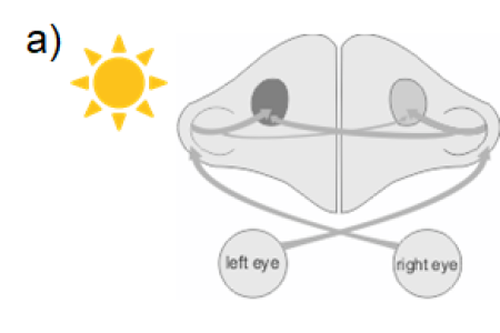

Hemispheric asymmetries represent a fundamental organizational principle of sensory, cognitive, or motor processing in the brains of many animal species. This lateralization is related to left-right differences in the structural organization of neural circuits, but how these asymmetries emerge during ontogeny is still poorly understood. A much discussed issue is the relative importance of genetic and environmental factors. In a current neuroanatomical study, biopsychologists from Bochum therefore investigated how projection asymmetries develop in the visual system of pigeons. Retrograde tracing of the major ascending (tectorotundal) projections in light-exposed and –deprived pigeons indicates that light stimulation during embryonic development leads to a stronger innervation of the left side of the brain. However, light does not enhance stabilization of fibers within the left hemisphere but induces stronger pruning of projections to the less stimulated right hemisphere. These data illustrate how visual input during early development modifies connectivity pattern in both brain halves, which in turn profoundly affects lateralized sensory processing, and ultimately lateralized cognitive processes, decision-making, or behavioral control.
Letzner S, Manns M, Güntürkün O. Light-dependent development of the tectorotundal projection in pigeons. Eur J Neurosci. 2020 Sep;52(6):3561-3571. doi: 10.1111/ejn.14775. https://onlinelibrary.wiley.com/doi/10.1111/ejn.14775
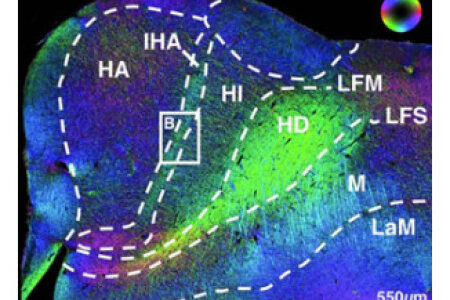

For more than a century, the avian forebrain has been a riddle for neuroscientists. Birds demonstrate exceptional cognitive abilities comparable to those of mammals, but their forebrain organization is radically different. Whereas mammalian cognition emerges from the canonical circuits of the six-layered neocortex, the avian forebrain seems to display a simple nuclear organization. Now, a new study headed by biopsychologists from Bochum, reveals a hitherto unknown neuroarchitecture of the avian sensory forebrain that is composed of iteratively organized cortex-like canonical circuits. This finding suggests that today’s birds and mammals use a partly modified ancient microcircuit of their last common ancestor. The avian version of this connectivity blueprint could conceivably generate computational properties akin to neocortex and would thus provide a neurobiological explanation for the comparable and outstanding perceptual and cognitive feats of both taxa.
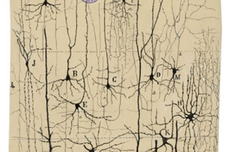

Neocortical neurons come in bewildering shapes and functions. Since more than a century neuroscientists have proposed hundreds of ways how to classify them, resulting in a jungle of contradictory nomenclatures. Now, 72 neuroscientists assembled in Copenhagen and proposed to start anew by classifying neocortical neurons based on single-cell transcriptomics that holds the promise of being complete, accurate, and suited to incorporate data from different approaches, developmental stages and species. Such a community-based classification could provide a common foundation for the study of cortical circuits.
Yuste, R., Hawrylycz, M., Aalling, N., Aguilar-Valles, A., Arendt, D., Arnedillo, R. A., Ascoli, G. A., Bielza, C., Bokharaie, V., Bergmann, T. B., Bystron, I., Capogna, M., Chang, Y., Clemens, A., de Kock, C. P. J., DeFelipe, J., Dos Santos, S. E., Dunville, K., Feldmeyer, D., Fiáth, R., Fishell,G. J., Foggetti, A., Gao, X., Ghaderi, P., Goriounova, N. A., Güntürkün, O., Hagihara, K., Hall, V. J., Helmstaedter, M., Herculano, S., Hilscher, M. M., Hirase, H., Hjerling-Leffler, J., Hodge, R., Huang, J., Huda, R., Khodosevich, K., Kiehn, O., Koch, H., Kuebler, E. S., Kühnemund, M., Larrañaga, P., Lelieveldt, B., Louth, E. L., Lui, J. H., Mansvelder, H. D., Marin, O., Martinez-Trujillo, J., Chameh, H. M., Nath, A., Nedergaard, M., Němec, P., Ofer, N., Pfisterer, U. G., Pontes, S., Redmond, W., Rossier, J., Sanes, J. R., Scheuermann, R., Serrano-Saiz, E., Steiger, J. F., Somogyi, P. S., Tamás, G., Tolias, A. S., Tosches, M. A., García, M. T., Vieira, H. M., Wozny, C., Wuttke, T. V., Yong, L., Yuan, J., Zeng H. and Lein, E., A community-based transcriptomics classification and nomenclature of neocortical cell types. Nature Neurosci., 2020, https://doi.org/10.1038/s41593-020-0685-8.
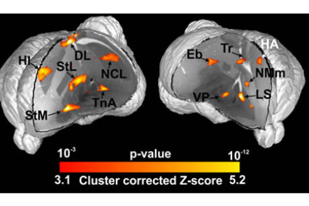

It is a long-time dream of avian neuroscientists to see the pattern of activity in a bird brain while the animal is working on a task. Now, biopsychologists and further colleagues from Bochum established an fMRI platform to investigate visually guided decision-making in awake behaving pigeons. Pigeons discriminated colors in a Go/NoGo paradigm and used their beak opening movements to signal their acceptance or rejection of the stimulus. This approach opens the door to visualize the neural fundaments of perceptual and cognitive functions in birds—a vertebrate class of which some clades are cognitively on par with primates.
Behroozi, M., Helluy, X., Ströckens, F., Meng, G., Pusch, R., Tabrik, S., Tegenthoff, M., Otto, T., Axmacher, N., Kumsta, R., Moser, D., Genc, E. and Güntürkün, O. (2020). Event-Related functional MRI in awake behaving pigeons. Nature Communications. 11, 4715. https://doi.org/10.1038/s41467-020-18437-1


Most humans demonstrably favor one limb over the other. Most research on limb preferences has been dedicated on handedness and its genetic foundation as well as its association with for example psychiatric disorders or language lateralization. However, humans also usually prefer one foot over the other. In this large-scale meta-analysis featuring over 145000 individuals, a multinational team from Germany, Greece and the UK aimed to conclusively determine (1) the prevalence of left- and right-footedness in the population, (2) the relationship between footedness and handedness and identify (3) moderating factors on footedness such as psychiatric disorders, being an experienced athlete and the individuals’s sex or age. We found that the prevalence of left footedness was 12.10% in the population. 60.1% of left‑handers were left‑footed, but only 3.2% of right‑handers were left ‑footed indicating a strong association between handedness and footedness. Furthermore, all above-mentioned moderators had a significant impact on the prevalence of footedness indicating that footedness is determined by both a nature and a nurture component. We hope that this study inspires more research on footedness as this phenotype has been critically understudied in laterality research.
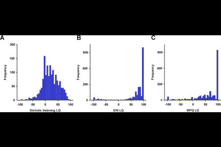

Humans display a large number of lateralized behaviors that are immediately visible such as handedness or footedness. These asymmetries are not limited to behavioral actions, but are also manifest in hemispheric lateralization of cognitive functions such as language or emotional processing. It is however unclear to what extent different asymmetries of brain and body correlate with one another. In this study, scientists from Bochum and Dresden investigated a large scale sample of over 1500 individuals with respect to their handedness, footedness and language lateralization using a dichotic listening task. We found almost no evidence that motor asymmetries and language lateralization were associated with one another using continuous lateralization quotients. On the contrary, we even found evidence that there is no link between these variables. These results indicate that previous studies suggesting an association between motor and cognitive hemispheric asymmetries might have overestimated their common biological and genetic basis.


Laterality research has come a long way since Pierre Paul Broca discovered differences between the left and right hemisphere in language lateralization. Since then, hemispheric asymmetries have been identified to be a fundamental principle in sensory, cognitive and motor processing alike indicating their paramount role in the information processing stream. In the past decade, neuroscientific research has developed a wide range of new methodological tools that significantly improve scientific insights and the reliability of findings in the field. However, laterality research has not yet embraced some of these new tools available to the community. In this perspective paper, we outline the 10 most outstanding challenges and opportunities in laterality research to us and how to approach each of them in the next decade. These issues encompass (1) the necessity for more meta-research, more representative samples and higher ecological validity to increase reproducibility and external validity, (2) using novel experimental and analysis techniques in both animal and human research and (3) exploring the role of laterality in psychiatric and neurodevelopmental disorders and their treatments. We hope that this paper inspires discussion and more research in the coming 10 years in laterality research to shape the next decade on a solid scientific fundament.
Ocklenburg, S., Berretz, G., Packheiser, J. and Friedrich, P. (2020): Laterality 2020: entering the next decade, Laterality, DOI:10.1080/1357650X.2020.1804396
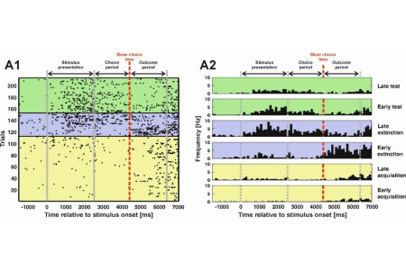

The ability to learn fundamentally rests on the prediction of events in the environment. If there is a mismatch between one’s expectations and the actual outcome, we are programmed to update our expectations based on reality. This process is known as reward prediction error-based learning and has successfully explained behavior in a large variety of species. On the neural level, the activity pattern of dopamine neurons in the midbrain has been identified to represent these reward prediction errors. However, only little is known about the precise temporal dynamics of reward prediction errors to this day. In our study, we investigated pigeons in an extinction learning paradigm that encompassed acquisition, extinction and a renewal test phase in a single session allowing for the investigation of single unit activity across learning. We found that neurons in the NCL, a structure strongly innervated by midbrain dopamine neurons, encode reward prediction errors both during the extinction phase as well as the renewal test. Gradually, the reward prediction error signal decreased with ongoing learning indicating the increasing match between expectations and reality. Furthermore, the neural signal transitioned from the outcome phase of the experiment to the presentation of the reward predictive cue. This is in line with temporal-difference models of learning. In total, our results show that reward prediction errors change gradually on a trial-by-trial basis during learning. Finally, we could demonstrate that reward prediction errors are truly expectancy-driven as we could elicit these signals without changing the quantity or quality of reward.
Packheiser, J., Donoso, J. R., Cheng, S., Güntürkün, O. and Pusch, R.. Progress in Neurobiology, https://doi.org/10.1016/j.pneurobio.2020.101901
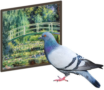

Visual categorisation is a useful way of organizing and simplifying incoming information from the envorinment around us. Over the past decade, birds have become a popular non-human animal model for vision due to their highly evolved visual systems and abilities. In this study we recorded neural activity from three pallial brain areas in pigeons: the nidopallium caudolaterale (NCL), an analogue of mammalian prefrontal cortex; the entopallium (ENTO), an intermediary visual area similar to primate extrastriate cortex; and the mesopallium ventrolaterale (MVL), a higher-order visual area similar to primate higher-order extrastriate cortex, while the birds performed an S+/S− Picasso versus Monet discrimination task. In NCL, we found that activity reflected reward-driven categorisation, with a strong left-hemisphere dominance. In ENTO, we found that activity reflected stimulus-driven categorisation, also with a strong left-hemisphere dominance. Finally, in MVL, we found that activity reflected stimulus-driven categorisation, but no hemispheric differences were apparent. We argue that while NCL and ENTO primarily use reward and stimulus information, respectively, to discriminate Picasso and Monet paintings, both areas are also capable of integrating the other type of information during categorisation. We also argue that MVL functions similarly to ENTO in that it uses stimulus information to discriminate paintings, although not in an identical way. The current study adds some preliminary evidence to previous literature which emphasises visual lateralisation during discrimination learning in pigeons.
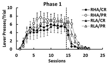

Individuals trained under partial reinforcement (PR) typically show a greater resistance to extinction than individuals exposed to continuous reinforcement (CR). This phenomenon is referred to as the PR extinction effect (PREE) and is interpreted as a consequence of uncertainty-induced frustration counterconditioning. In this study, we assessed the effects of PR and CR in acquisition and extinction in two strains of rats, the inbred Roman high- and low-avoidance (RHA and RLA, respectively) rats. These two strains mainly differ in the expression of anxiety, the RLA rats showing more anxiety-related behaviors (hence, more sensitive to frustration) than the RHA rats. At a neurobiological level, mild stress is known to elevate corticosterone in RLA rats and dopamine in RHA rats. We tested four groups of rats (RHA/CR, RHA/PR, RLA/CR, and RLA/PR) in two successive acquisition-extinction phases to try to consolidate the behavioral effects. Animals received training in a Pavlovian autoshaping procedure with retractable levers as the conditioned stimulus, food pellets as the unconditioned stimulus, and lever presses as the conditioned response. In Phase 1, we observed a PREE in lever pressing in both strains, but this effect was larger and longer lasting in RHA/PR than in RLA/PR rats. In Phase 2, reacquisition was fast and the PREE persisted in both strains, although the two PR groups no longer differed in lever pressing. The results are discussed in terms of frustration theory and of uncertainty-induced sensitization of dopaminergic neurons.


Brain asymmetries are key components of sensory, cognitive, and motor systems of humans and other animals. This comprehensive review from Bochum biopsychologists tracks their development from embryological asymmetries of genetic expression patterns up to left-right differences of neural networks in adults. It also offers on overview of the evolution of neural asymmetries as well as the history of its discoveries. These insights are crucial to understand the functional pattern of the lateralized human brain.


There is a long history of research linking lateralization to cognitive abilities, but so far this association had not been investigated in cats. An international research team from Turkey, Portugal and Germany of which the biopsychology was a part now investigated the association between hemispheric asymmetries and cognitive in cats. To this end, strength and direction of paw preferences in 41 cats were measured using two novel food reaching tasks in which the animals needed to open a lid in order to reach the food reward. The research team found that cats that showed a clear preference for one paw were able to open more lids succesfully than ambilateral animals. Moreover, cats that preferred to interact with the test apparatus with their paw from the beginning, opened more lids than cats the first tried to interact with the test apparatus using their heads. Results also suggested a predictive validity of the first paw usage for general paw usage. It was also shown that the cats' individual paw preferences were stable and task-independent. These results yield further support to the idea that lateralization may enhance cognitive abilities.
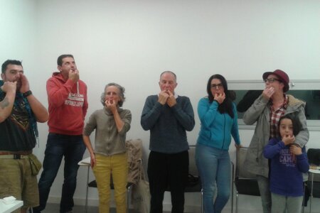

Silbo Gomero is a Spanish whistled language that is mainly used on the island of Silbo Gomero. It has rarely been investigated neuroscientifically, but a research team from the biopsychology now analyzed lateralization in this unusual language. The research team used a vocal and a whistled dichotic listening task in a sample of 75 healthy Spanish speakers. Both individuals that were able to whistle and to understand Silbo Gomero and a non-whistling control group showed a clear right-ear advantage for vocal dichotic listening. For whistled dichotic listening, the control group did not show any hemispheric asymmetries. In contrast, the whistlers’ group showed a right-ear advantage for whistled stimuli. This right-ear advantage was, however, smaller compared to the right-ear advantage found for vocal dichotic listening. In line with a previous study on language lateralization of whistled Turkish, these findings suggest that whistled language processing is associated with a decrease in left and a relative increase in right hemispheric processing. This shows that bihemispheric processing of whistled language stimuli occurs independent of language.


How many people really are left-handed? An international research team from Greece, the UK and Germany of which the biopsychology lab was a part now conducted the largest meta-analysis in the world on left-handedness to clarify this question. The researchers could show that prevalence of left-handedness lies between 9.3% to 18.1%, with the best overall estimate being 10.6%. Handedness variability depends on (a) study characteristics, namely year of publication and ways to measure and classify handedness, and (b) participant characteristics, namely sex and ancestry.


A significant aspect of human social interaction revolves around touching another individual. The most common social touch behaviors are embracing, kissing and the cradling of children, all of which have been demonstrated to be lateralized on the population level in humans. Two major theories have been proposed driving this type of laterality, namely via motor preferences or thorugh an emotional bias. In this study, we systematically tested predictions of both theories as participants had to perform all three social interactions with inanimate objects such as dolls or mannequins in neutral, positive or negative emotional situations. We found that biases in kissing and cradling were unaffected by the handedness of the participants. Only embracing laterality was significantly correlated with handedness. Furthermore, we found that lateralization was reduced in emotional compared to neutral conditions indicating that affective states influence the lateralization of human social touch. Overall, these results thus support the notion that lateral biases in social touch are driven by emotive rather than motor biases.

Since the days of Pavlov, associative learning has become a powerful tool to explain behavior of a large variety of organisms. One of the most prominent models of associative learning is the Rescorla-Wagner model. It assumes that different cues massively compete for associative strength. However, some studies have shown that predictions of the Rescorla-Wagner model fail under certain circumstances. Recently, it has been argued that failures of the model result from the systematic overestimation of the extent of cue competition. In our study, we aimed to test different levels of cue competition using an overexpectation design in which cues are being extinguished despite continuous presentations of reward. In a first experiment, we found no evidence for cue competition. For this reason, an additivity training was introduced into the second experiment to strengthen the level of cue competition. In the second experiment, we were able to demonstrate cue competition, however only to a moderate rather than a massive extent as is predicted by the Rescorla-Wagner model. Overall, these results show that cue competition can be modulated and that its extent indeed seems to be slightly overestimated by the Rescorla-Wagner model.
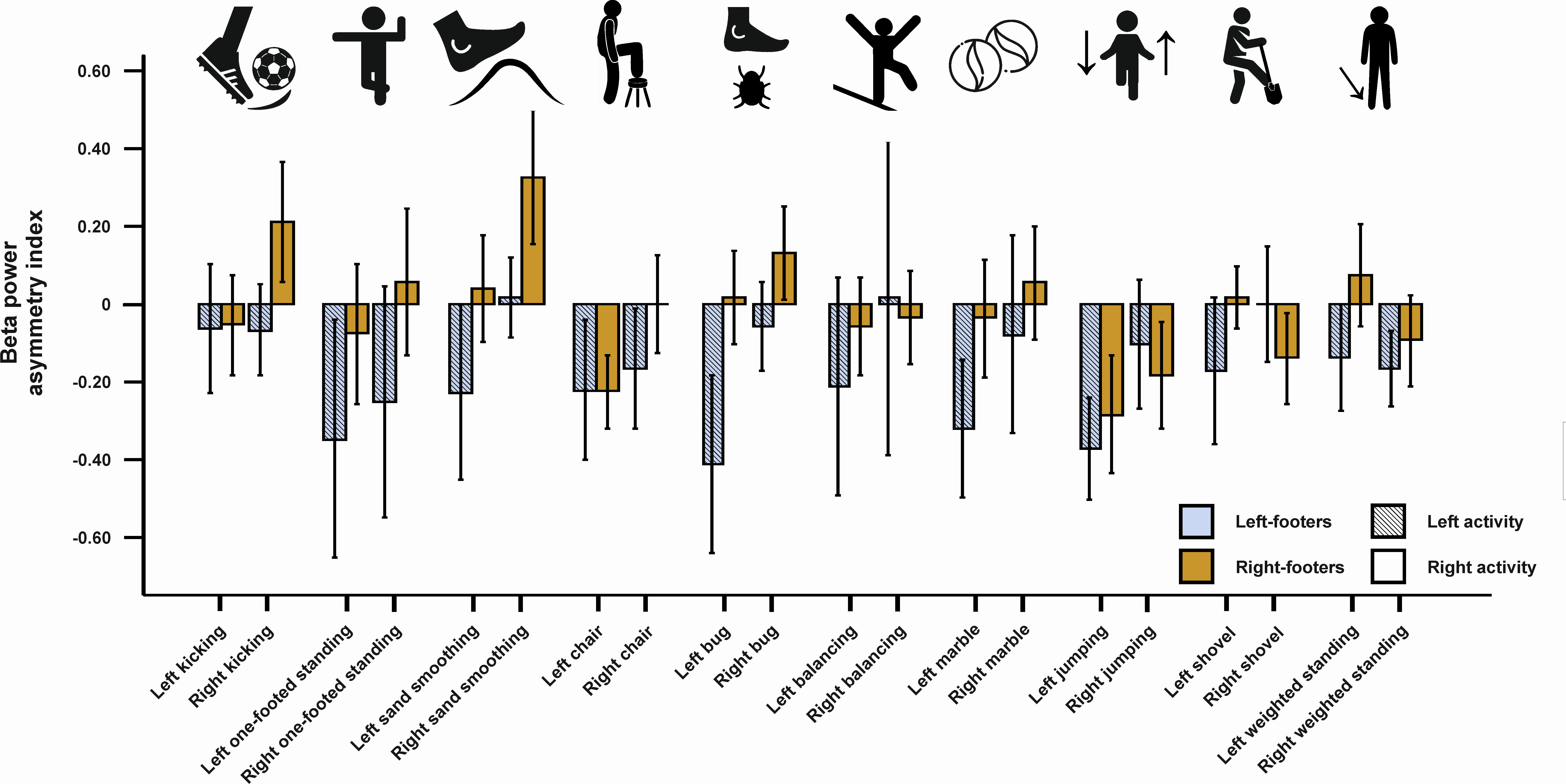

The neural basis of motor asymmetries such as handedness and footedness has been studied widely using both EEG and fMRI studies in humans. However, these studies almost exclusively used both unnatural tasks such as finger tapping and unnatural environments such as an fMRI scanner to identify the underlying biological correlates of motor functions. To overcome these shortcomings, our study investigated handedness and footedness in ecologically valid settings by using a mobile EEG in which participants could freely move around. We then let them perform tasks as described by the Edinburgh Handedness Inventory (EHI) and the Waterloo Footedness Questionnaire (WFQ), two of the most widespread questionnaires to assess motor asymmetries. Thus, our participants were shooting and throwing balls, balancing on rails and even jumped around while being recorded by an EEG. We found that both alpha and beta oscillations on fronto-central electrodes differed significantly between left- and right-handers as well as left- and right-footers. Furthermore, we could predict the extent of handedness and footedness using the EEG recordings during tasks performed with the left limb. All of these results were unaffected by movement indicating that mobile EEGs provide a powerful tool to study motor asymmetries in the field and can thus give meaningful insights into the neural correlates of real-life behavior.
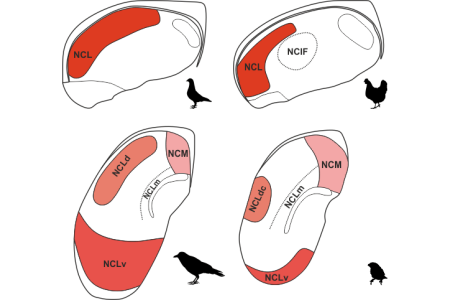

Approximately 40 years ago, Divac and colleagues first described the analogue to the prefrontal cortex (PFC) in birds known as the nidopallium caudolaterale (NCL). The NCL is comparable to the PFC as that both are seen as the seat of complex behaviors such as working memory and decision making. The past decades of research also demonstrated striking parallels in the underlying connectivity and physiology. Up until now, the location of the NCL had been described in pigeon only, and we knew close to nothing about the borders of the NCL in other bird species. This is problematic because there is considerable variation in both brain structure and behavioral capacities between different bird species. By visualizing the dopaminergic innervation of the forebrain, which is a hall-mark method to identify both the NCL and the PFC, a team of the Biopsychology lab set out to delineate the NCL in chicken, zebra finch and the carrion crow. They found that whereas the trajectory of the NCL in chicken is very similar to pigeon, the zebra finch and crow show a strikingly different pattern. In these two songbirds, the NCL now spans across the entire back of the forebrain and seems split up in to more distinct sub-units. These findings commensurate to what has been observed in mammals; the PFC in chimpanzees is both larger and more parcellated compared to rats. Thus, this study discloses yet another instance of the remarkable parallel evolution of the executive brain structure in birds and mammals.
von Eugen, K., Tabrik, S., Güntürkün, O. and Ströckens, F. (2020). A comparative analysis of the dopaminergic innervation of the executive caudal nidopallium in pigeon, chicken, zebra finch, and carrion crow. Journal of Comparative Neurology. https://doi.org/10.1002/cne.24878


Those who were humiliated, beaten or sexually abused in childhood have to deal with mental illnesses such as depression or anxiety attacks more often in adulthood than people who were spared this experience at a young age. But what are the reasons for this greater vulnerability? Do experiences of violence as a child possibly lead to a permanently changed perception of social stimuli? Psychiatrists from Bonn and biopsychologists from Bochum now indeed were able to shed light on basic factors relevant to social dysfunction, interpersonal distance, and responsiveness to touch in adults with a history of childhood abuse. In addition to behavioral testing, structural and functional MRI was performed to assess the neural correlates of social touch in relation to varying degrees of maltreatment. Overall, the findings suggest alterations in neural responsivity to sensory stimulation with both fast and slow touch. Of particular interest was the finding that severe maltreatment was associated with decreased hippocampal responsivity to slow touch, which tends to be associated with the emotion-relevant, comforting aspects of touch. In addition, the scientists also examined social distance by asking participants to approach an unknown person and to stop when the distance was just felt to be pleasant. Indeed, traumatized subjects kept a larger distance by twelve centimeters. These findings upon up new possibilities for body-based therapies of traumatized subjects.

Corvids, parrots and other bird groups demonstrate complex cognition, including causal reasoning, mental flexibility, planning, social cognition and imagination. These cognitive abilities were a surprise to many scientists. They were not expected to be found in birds because of their small brains and the absence of a cerebral cortex. Onur Güntürkün now explains in Scientific American that it is possibly less of interest that both birds and mammals succeeded in growing smart. Rather this accomplishment came about through development of mostly identical neural mechanisms despite differently organized forebrains. Birds and mammals cognitively thrived by increasing neuron numbers. Mammals did so by expanding brain size and birds by amplifying neuron density. They both developed substantially similar networks of “cortical” connections and evolved “prefrontal” areas with identical physiological, neurochemical and functional features. The same can be said for cognition itself. The way birds and mammals learn, remember, forget, err, generalize and make decisions follows identical principles. This astonishing degree of similarity is only possible when nature offers severely limited degrees of freedom in generating neural structures for complex cognition. Birds and mammals evolved similar neural mechanisms and ways of thinking— taking different paths that ended in the same place.
Güntürkün, O., The surprising power of the avian mind, Scientific American, 2020, January, 48-55.
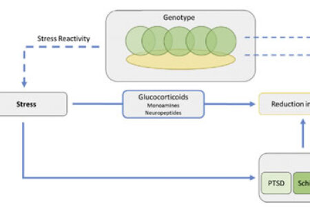

In healthy humans, the brain shows typical differences between the left and the right hemisphere in structure and function – so-called hemispheric asymmetries. In different psychiatric and neurodevelopmental disorders like schizophrenia, autism spectrum disorder, dyslexia, depression and posttraumatic stress disorder however, many asymmetries are reduced. Most of these disorders show a small genetic overlap with each other but none with asymmetry-related genes. A common non-genetic factor among these disorders are changes in the stress system as well as intra- and early life stress. A team from the Biopsychology and the Cognitive Psychology lab from the Ruhr University Bochum now propose a model in which early life stress as well as chronic stress not only increases the risk for psychiatric and neurodevelopmental disorders but also changes structural and functional hemispheric asymmetries. This influence might arise from prenatal effects of maternal stress and early life stress on the stress system and disease development: maternal adversity influences the environment in the womb which in turn programs neural systems underlying cognitive-emotional function. This could also be the case for the development of hemispheric asymmetries. Moreover, pathological changes in the stress system as well as hemispheric asymmetries may lead to further dysregulation in the brain. However, the effect of stress on hemispheric asymmetries could depend on the timing and type of the stressor.
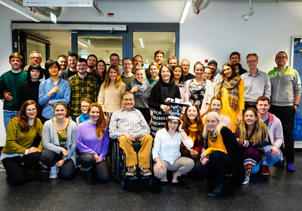

On Thursday, December 12, 2019, Caro successfully defended her doctoral thesis on “Personality Neuroscience: Biological Basis of Neuroticism and Volition”.
If one types “Personality Psychology” into a search engine and just opens the first Wikipedia link, the astonishing finding is that this branch of psychology is so remote from neuroscience as hardly any other field of psychological inquiry. As beautifully laid out in Caro’s introduction to her thesis, this is a bit related to the failure of the first grand neuropsychological theory of Eysenck. But it is also a result of serious methodological and conceptual problems. Caro bravely steps into this missing link by focusing on neuroticism and volition. This last point implies also a reach-out into motivation research. In a series of breath-taking studies, Caro then demonstrates links between the dendritic organization of parts of the amygdala and neuroticism, the impact of efficient dopaminergic transmission and the tendency to procrastinate, and correlations between types of action orientation and gender as well as intelligence. Most importantly, she also demonstrates that not only amygdala volume but also the functional connectivity between amygdala and the anterior cingulum is correlated with the tendency of individuals to either start acting or to go on waiting. This last study was published in Psychological Science and became a media bestseller. Each and every of these studies is written by Caro with a profound knowledge of psychology and neuroscience and with the desire to achieve mechanistic explanations. Thus, Caro demonstrates her ability to be a perfect biosychologist.
The committee consisting of Onur Güntürkün, Nikolai Axmacher, Boris Suchan, and Robert Kumsta was deeply impressed and awarded this great thesis with a magna cum laude. Soon after this success, Caro fulfilled her plan to leave science, but with the promise to hopefully come back.
Congratulations Caro for being such an outstanding biopsychologist! We terribly will miss you.


On Wednesday, the 4th of December 2019, Steffi successfully defended her doctoral thesis on the “Mechanisms of light-induced neuronal asymmetry during embryonic development” at the IGSN. Most vertebrate brains display neuro-functional asymmetries and we know very little about their ontogeny. Steffi used in her PhD the visual asymmetry of pigeons to study the impact of microRNAs on asymmetry development. MicroRNAs are small non-coding RNAs that regulate many downstream biological processes. Although very promising, close to nothing is known about that in pigeons. Despite this challenge, Steffi managed to discover the critical main events in a tour de force of newly established techniques. First, she established from scratch microRNA analysis in our lab and demonstrated that there is no asymmetry in the retina but in the tectum for embryonic expression of the microRNA-183 cluster. To move deeper, she established qPCR and showed that all constituent parts of the microRNA-183 cluster in the right tectum were low when dark, but very high when light incubated. The left tectum evinced only intermediate expression levels after light incubation. Thus, light seems to be needed to induce expression but then primarily activates the not-light exposed right tectum! Puzzling. To solve this, Steffi reconstructed the hottest candidates for the targets of microRNA-183 and found pathways for growth and signal transduction. Then she started to identify with in situ hybridization and laser microdissection the anatomical localization of neurons that are modified by miR-183. Here, she discovered upper and lower tectal cells differ between left and right with respect to the different sub groups of microRNA-183 stained cells. Like Sherlock Holmes, she then puts the puzzle together and puts forward a likely scenario of molecular and cellular events that define the first steps of an asymmetrical brain. The committee consisting of Onur Güntürkün, Carsten Theiß, Giorgio Vallortigara, Thorsten Müller, and Laurenz Wiskott was deeply impressed and awarded this grand work with a summa cum laude.
Congratulations Steffi! We are extremely proud of you.


On Friday, October 25, 2019, Meng successfully defended her doctoral thesis on the “Neural circuits of appetitive extinction in the pigeon brain”. Extinction learning is a key learning procedure that proceeds with predictable sequences of behavioral changes in all studied vertebrates and invertebrates. But do these similarities of behavior also point to equivalencies of neural mechanisms? This is the core question of Meng’s thesis. She approaches it by behavioral pharmacology and fMRI. In her behavioral studies she probes the contribution of a visual associative structures and can show that it contributes to the processing of the context in learning extinction. This is highly interesting since context memory is in mammals the contribution of the hippocampus. Previous studies could not convincingly show that the pigeon’s hippocampus has a truly visible contribution to memorize the extinction context. Then Meng moves on to study the medial striatum and demonstrates that its contribution is mostly in the realm of the acquisition of extinction, possibly due to insufficient prediction error coding. Finally, she moves on to study for the first time extinction learning in a scanner and yields a huge amount of novel insights on the extinction network. Among these novel information is the finding that the first extinction session is characterized by a broad activation of a large number of forebrain areas. With every further extinction session, however, these activations get smaller and smaller and astonishingly encompass primary visual areas. Based on all these new data, Meng sketches the outline as well as the details of the avian extinction network. She shows that its main outline is similar but not identical to that of mammals. The committee consisting of Onur Güntürkün, Nikolai Axmacher, Martina Manns, and Maik Stüttgen was deeply impressed and awarded this great series of studies with a magna cum laude.
Congratulations Meng! You are fantastic.

On Thursday, the 24th of October 2019, Christoph successfully defended his doctoral thesis on the “Neural Correlates of Intelligence and General Knowledge Derived by Magnetic Resonance Imaging”. Christoph’s thesis is in many ways unique. While there are several studies on the neural foundations of fluid intelligence, there is practically no such study on the brain fundaments of crystallized intelligence. Christoph’s thesis introduces the reader with a fantastic clarity through the history and present status of the psychology and the neuroscience of intelligence research, to then develop his own way of studying this field. In 324 (yes: 324!) subjects he collects state-of-the-art functional and imaging data and scrutinizes the knowledge base and fluid intelligence of his subjects. He can show that fluid intelligence is equal in both sexes, but that men churn out their fluid intelligence by grey matter volume, while women do it with a highly efficient functional connectivity. For crystallized intelligence, however, women score lower than men, due to differences in structural connectivity. In the next study he uses NODDI to reconstruct the cellular geometry of grey matter and can show that dendritic morphology that derives from efficient synaptic pruning correlates with IQ. This study, that was published in Nature Communications, was among the 0.02% most publicly discussed scientific papers of the last years. Finally, Christoph reconstructs the connectional fundaments of fluid intelligence by the using the functional connectome of the human brain. The committee consisting of Onur Güntürkün, Nikolai Axmacher, Oliver Wolf, and Boris Suchan was deeply impressed and awarded this grand work with a magna cum laude.
Congratulations Christoph! We are very proud of you.
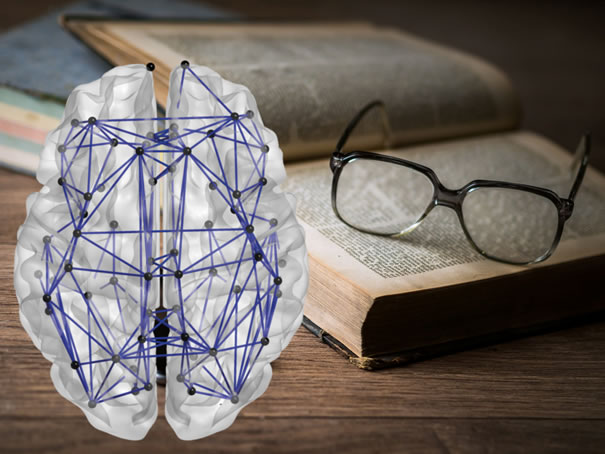

What is the only constant in the theory of relativity? Can you name the capital of Sweden? Who wrote "The Divine Comedy"? Some individuals are more capable of answering such questions than others. Our knowledge about the world around us makes us who we are, and profound general knowledge can help us navigate through everyday life unerringly. The individual pieces of information constituting our general knowledge are likely to be encoded on a microstructural level, namely within the myriads of synapses of our brain. This gives rise to the question whether interindividual differences in general knowledge can also be related to macrostructural differences in the biological properties of the brain. Now, for the first time, a team of scientists from Bochum and Berlin has examined the neural correlates of general knowledge. They reported their results in "European Journal of Personality".
Together with colleagues from Ruhr University Bochum and Humboldt University Berlin, Dr. Erhan Genç examined the brains of 324 men and women using diffusion tensor imaging. This enabled the scientists to reconstruct the trajectories of nerve fibers in the brain and to gain insight into its structural network. Graph theoretical computations allowed the researchers to assign an individual value to each of the participants' brains reflecting the efficiency of this structural network. In addition to MRI measurements, participants also completed a general knowledge questionnaire developed in Bochum, the so-called Bochumer Wissenstest. This test included more than 300 questions from various fields of general knowledge, such as art and architecture or biology, physics and chemistry. Finally, Dr. Erhan Genç and his colleagues investigated whether the efficiency of the brain's structural network was associated with the amount of general knowledge a person holds.
The scientists were able to show that individuals with a very efficient fiber network actually had more general knowledge than those with a less efficient network. Importantly, this association was not driven by other factors such as age or sex. Given that general knowledge is likely to be stored in the form of single information fragments scattered across the whole brain, it is conceivable that an efficiently organized network of nerve fibers could provide a beneficial infrastructure for the storage of new information. In the long run, this might foster general knowledge acquisition and lead to higher results in respective test instruments.


Everyone knows that about 90% of humans are right-handers and 10% are left-handers, but did you know that animals also hava preference to use one paw over the other? In the present study and international team of researchers from Bochum, Turkey and Greece statistically integrated findings on paw preferences in cats and dogs, two species are commonly kept as pets in many societies around the globe. For both species, there were significantly more lateralized than non-lateralized animals. We found that 78% of cats and 68% of dogs showed either left- or right-sided paw preference. Unlike humans, neither dogs nor cats showed a rightward paw preference on the population level. For cats, but not dogs, we found a significant sex difference, with female animals having greater odds of being right-lateralized compared to male animals.
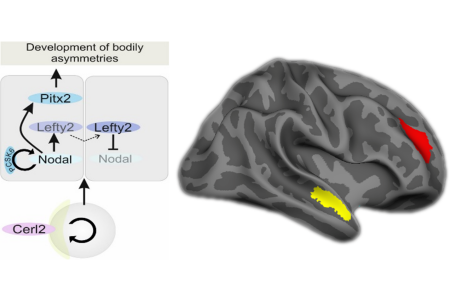

From the outside, the human body seems symmetrical. On the inside however, organs are arranged asymmetrically - the heart for example has its seat on the left side in most cases. The development of bodily asymmetries is influenced by the nodal cascade. Here, the gene PCSK6 has shown regulatory function during left-right axis formation by initiating symmetry breakage. However, it is not clear if variation in this gene is also associated with structural asymmetries in the brain. To examine the impact of PCSK6, biopsychologists from the Ruhr-University Bochum analyzed the effects of genetic variation in PCSK6 in a cohort of 320 healthy adults. They linked different PCSK6 variants to the asymmetry in the superior temporal sulcus, a structure involved in voice perception. Heterozygous individuals who carry a short and a long PCSK6 allele showed stronger rightward asymmetry. Further associations were evident in the dorsolateral prefrontal cortex. Here, individuals homozygous for short alleles showed a more pronounced asymmetry. This indicates that PCSK6, a gene that has been implicated in the ontogenesis of bodily asymmetries by regulating the nodal cascade, is also relevant for structural asymmetries in the human brain.


The neurocognitive research building THINK (Zentrum für Theoretische und Integrative Neuro- und Kognitionswissenschaft) is now finally approved by the Joint Science Commission that is constituted by the German Ministry of Science and Education as well as the local science ministries of all Federal States of Germany. The commission thus follows the advice of the Wissenschaftsrat (Central Science Commission of Germany). THINK was even chosen as the best science building application of 2019 and will be funded with 89 million Euro.
This new center will be the future core point of research on the neuronal mechanisms of cognition, hybrid cognitive systems, and artificial intelligence in Bochum. It is the first German science building ever that was successfully applied from a speaker from psychology (Onur Güntürkün) and will house ca. 100 scientists who can run within 4,000m2 cutting edge research that span from philosophy of mind to single cell recording, and from technical applications to high field imaging. THINK is planned to be ready in 2024 and will be located on the north edge of the previous Opel car plant.


Some people tend to postpone tasks, although being aware of the adverse effects accompanying procrastination. Our individual ability to tackle tasks directly instead of putting them off is highly dependent on our ability to initiate cognitive-, motivational- and emotional-control mechanisms, so-called metacontrol. Even though individual differences in these metacontrol mechanisms have far-reaching influence our personal and professional life, their genetic foundation is somewhat unknown. Biopsychologists and human geneticists from the Ruhr University Bochum and the University of Technology Dresden joined forces to explore the genetic foundations of interindividual differences in trait-like procrastination, measured as decision-related action control (AOD). The researchers investigated the individual AOD score as well as the genotype of 278 healthy adults. Here they were particularly interested in the tyrosine hydroxylase gene (TH gene). The expression of the TH gene influences the availability of neurotransmitters from the catecholamine family, including dopamine. Since dopaminergic signaling plays a significant role in various metacontrol processes, they aimed to examine whether genetically induced differences in the dopaminergic system are associated with interindividual differences in AOD. The analysis yielded a sex-dependent effect of TH genotype on AOD.
Interestingly, only in women, a genetic predisposition towards higher dopamine levels was associated with poorer action control and therefore the trait-like tendency to procrastinate. Additionally, the research group investigated whether differences in the morphology and functional connectivity of the amygdala that were previously associated with AOD happen to be related to differences in the TH genotype and thus to differences in the dopaminergic system. However, there was no significant amygdala volume or connectivity difference between the TH genotype groups. Therefore, this study is the first to suggest that genetic, anatomical, and functional differences affect trait-like procrastination independently.
Schlüter C., Arning L., Fraenz C., Friedrich P., Pinnow M., Güntürkün O., Beste C., Ocklenburg S. & Genc E.. Genetic variation in dopamine availability modulates the self-reported level of action control in a sex-dependent manner. Social Cognitive and Affective Neuroscience (2019) 1–10. https://doi.org/10.1093/scan/nsz049


Have you ever noticed that children are mostly carried on the left body side of the parent? This lateral preference in infant cradling has been scientifically investigated for 60 years, but influential factors underlying this bias have remained largely unknown due to inconsistent experimental designs and low sample sizes. Luckily, meta-analyses resolve such issues, so we analyzed the entire body of literatur on this topic and generated three major findings on the cradling bias: (1) approximately 69% of all adults prefer to cradle children on the left. (2) Right-handed people have a stronger left-sided preferences than left-handed people. And (3), women have a stronger left-sided preference compared to men. These findings complement recent hypotheses on motor and emotional biases on the laterality of social touch in humans. And if you find yourself holding a baby next time, pay attention to your holding side. It might just tell you something about your feelings!

No matter whether it is in sports or academics, we need to overcome hurdles to reach our goals. In this context, we often ask ourselves why some people get along more successfully than others and where the differences are between those who stumble and those who do not. Individual differences in the ability to initiate self- and emotional-control mechanisms have been related to personal and professional success repeatedly. These differences have been explicitly described in Kuhl’s action-control theory. Although interindividual differences in action control make a significant contribution to our everyday life, their neural foundation remains unknown. To gain deeper insight into the neuronal basics of action control, biopsychologists from Bochum measured action control in a sample of 264 healthy adults and related interindividual differences in action control to variations in brain structure and resting-state connectivity. Their results demonstrate a significant negative correlation between decision-related action orientation (AOD) and amygdala volume. Thus, individuals with larger amygdala volume were less successful to use self- and emotional control mechanisms to achieve a particular goal and in turn, more prone to procrastination and hesitation. Further, the functional resting-state connectivity between the amygdala and the dorsal anterior cingulate cortex (dACC) was significantly associated with AOD. Here less functional connectivity was associated with lower AOD scores. Altered resting-state connectivity between these two brain areas might affect the top-down regulation between the dACC and the amygdala, that is crucial for successful action control. These findings are the first to show that interindividual differences in action control, namely AOD, are based on the anatomical architecture and functional network of the amygdala.
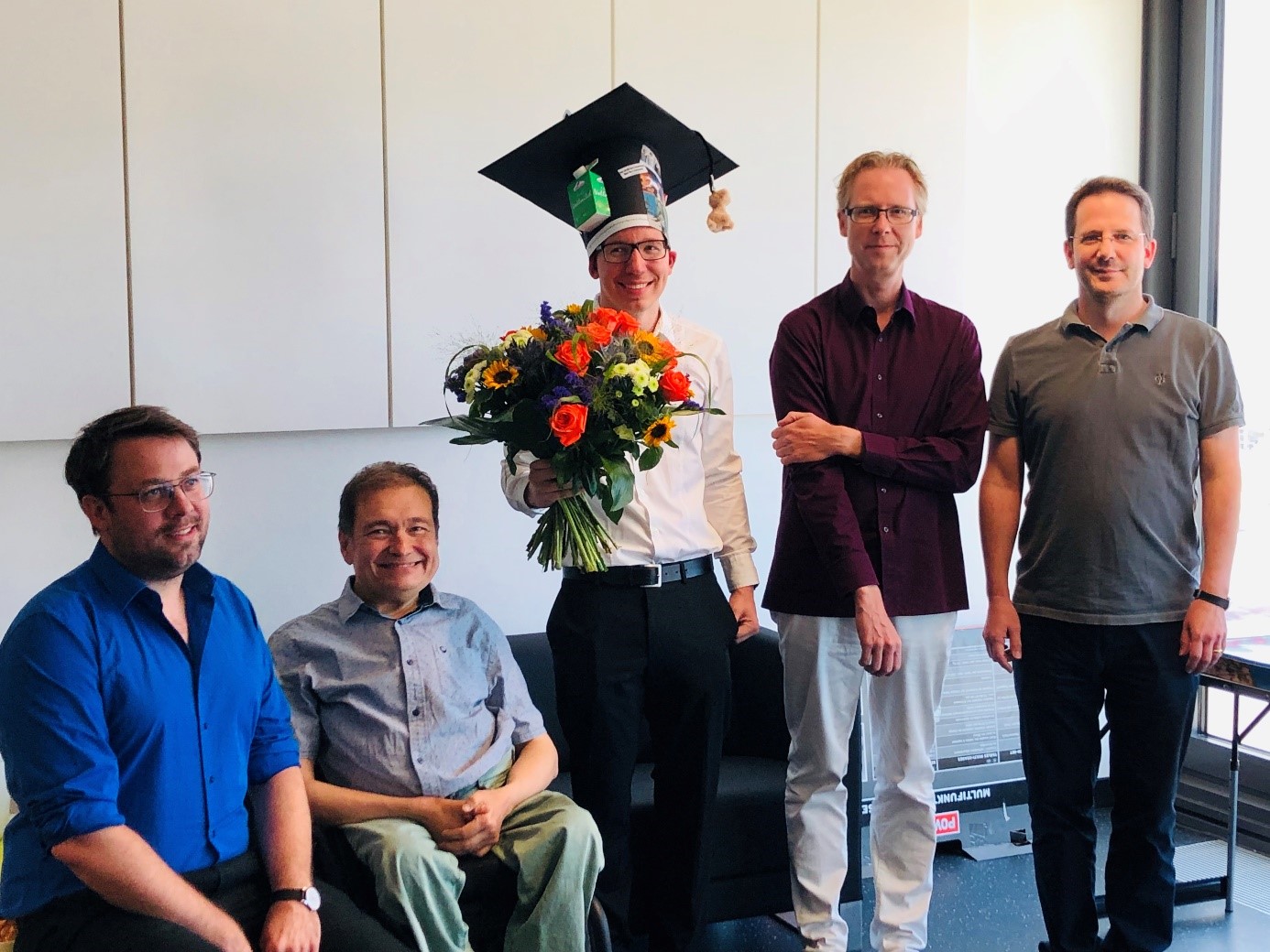

On Friday, the 28th of June 2018, Julian successfully defended his PhD thesis entitled "The neuronal mechanisms and dynamics of extinction learning and renewal". Julian’s PhD thesis puts two bold hypotheses forward: First, consolidation of extinction is not required for extinction to occur. Julian was quite cautious to also remark that this is true for behavior analyses and must not necessarily apply for all aspects of neurobiology. Second, key events during extinction are not coded by highly specialized neurons but by cells of mixed-selectivity as they encode a large variety of parameters during an extinction learning paradigm. While the neural responses are highly distributed and diverse on the single unit level, the population response can be decoded into discrete neural archetypes. These and many more findings were pretty impressive. Even more impressive was Julian’s ability to then successfully master 90 minutes of savage grilling by Onur Güntürkün, Nikolai Axmacher, Oliver Wolf, and Jonas Rose. The committee was so impressed that they decided to award Julian’s PhD with a summa cum laude. Congratulations, Julian! We all are very proud of you! During the subsequent photo session three people took in parallel pictures from different locations. The result is funny: everybody looks somewhere else.

Probably, none of us has ever experienced a serious and long-term shortage of food. But in principle evolution has endowed us and other vertebrates with mechanisms that alter our physiology, brain mechanisms, mental states, and behavior when environmental cues signal an upcoming scarcity of food. But what are these mechanisms? Biopsychologists now published a comprehensive theory on the mechanisms of foraging in the top tier theory-journal “Behavioral and Brain Sciences”.
The theory departs from the observation that both mammals and birds are extremely sensitive to a drop in the probability to find food and reliably react with an invigoration of food-related responses. During such a time these animals consume and/or hoard more food, get fatter, and build up a reserve against future starvation. This even happens when people are anxious to lose their job. In this paper it is argued that an anticipated long-term drop of resources has motivational effects called incentive hope that facilitates foraging effort by an altered release of dopamine - a neurotransmitter strongly involved in inducing incentive motivation. This increases the subjective value of food, promotes higher efforts to collect and consume it, and results in higher bodily or hoarded long-term energy storages. The paper shows that this hypothesis is computationally tenable, leading foragers in an unpredictable environment to search for and to consume more food items. Altogether, this theory bridges the troubled waters between ecology, psychology, and neurobiology. Twenty-three research groups have responded to the call to discuss this novel theory. Their comments are subsequently discussed and integrated into the body of their theory.
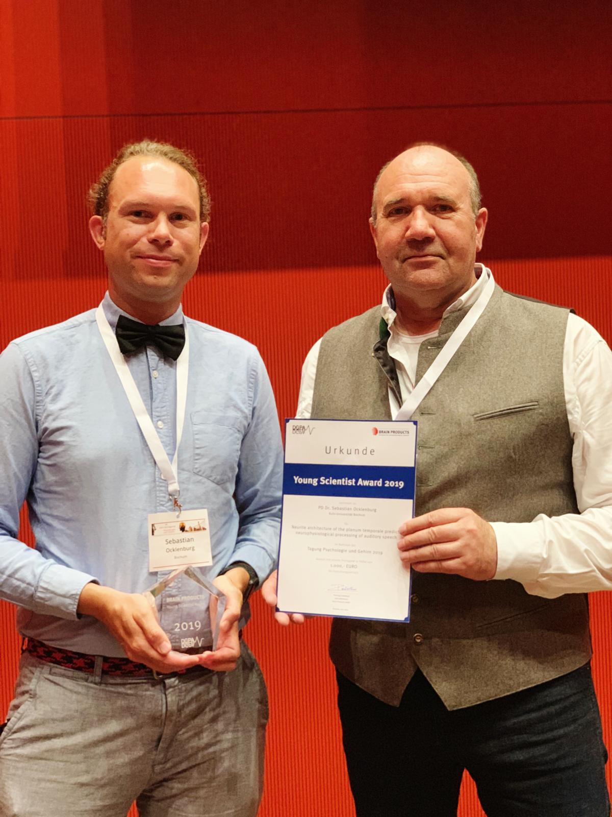

Sebastian Ocklenburg won the Brain Products Young Investigator Award 2019. This prestigous young investigator award was given to Sebastian Ocklenburg in an award ceremony during the "Psychology and Brain 2019" (Psychologie und Gehirn 2019) conference in Dresden for his work on language lateralization.
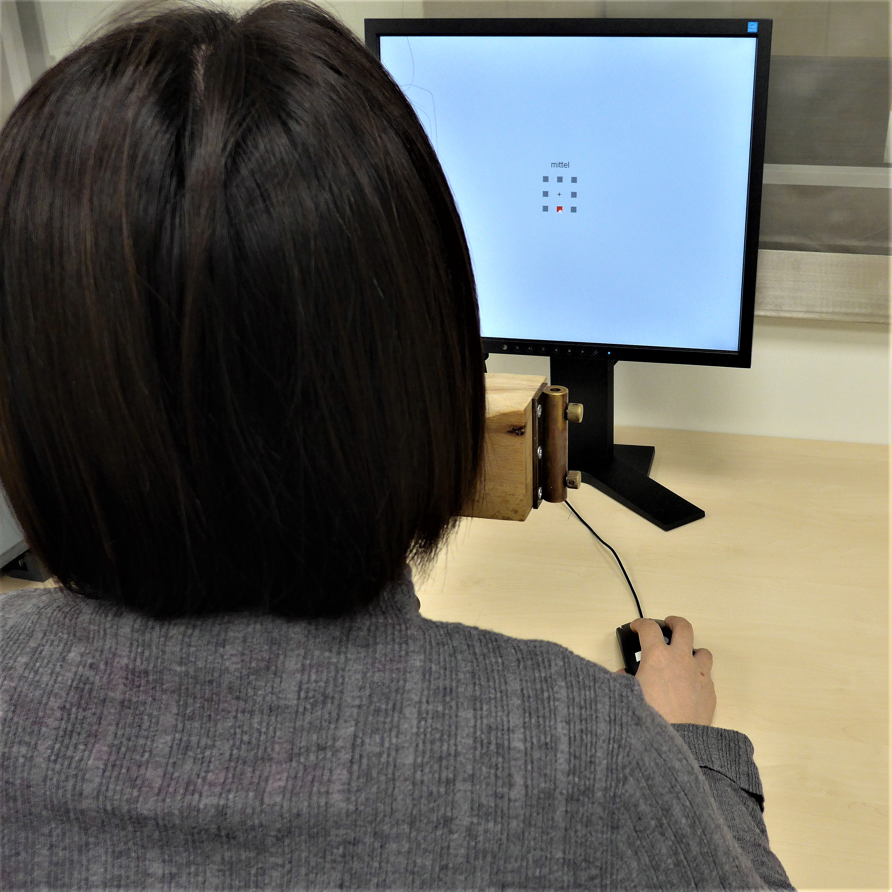

Among functional hemispheric asymmetries in humans, handedness is by far the most investigated. However, the underlying neural correlates remain unclear. Previous research has mainly focused on functional imaging methods and suggests differences in ipsilateral activation during unilateral hand movements between left- and right-handers. As EEG allows for a higher temporal resolution, researchers from the Biopsychology lab adapted the classical Tapley and Bryden task for use during EEG and tested this paradigm in 36 left- and 36 right-handers. Subjects had to click as fast and accurate as possible on eight squares distributed around a fixation cross in a given sequence with varying complexity. The lateralized readiness potential (LRP) is an event related potential component related to asymmetric motor preparation. Increasing complexity of sequences was associated with earlier and less negative LRP peaks. For the last response within a sequence, right-handers had more negative LRP peak amplitudes than left-handers. The effect of handedness on LRP peak amplitude in the first response was modulated by task complexity with a more negative LRP peak amplitude in right-handers than left-handers in simple, but not in medium or complex trials. This effect might be due to more symmetrical processing in right-handers with increasing task complexity, which complements findings from previous imaging studies.
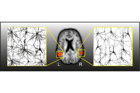

Post mortem studies have found hemispheric asymmetries in microstructure in several areas of the human brain. However, until recently it was impossible to assess microstructural asymmetries in vivo. In cooperation with the Department of Neurosurgery from Unviersity of New Mexico, a group of researchers from the Biopsychology lab used neurite orientation dispersion and density imaging (NODDI) to examine microstructural asymmetries in more than 500 participants to determine if findings are in accordance with what has been reported in post mortem studies. Greater left- than right-hemispheric estimated neurite density was found in early auditory, inferior parietal and temporal-parietal-occipital areas. In contrast, greater right-hemispheric neurite density was found in the fusiform and inferior temporal gyrus, reflecting what has been reported in post mortem studies. Microstructural asymmetries were mostly independent from participants’ sex or handedness. These findings suggest substantial microstructural asymmetries in gray matter, making NODDI a promising marker for future genetic and behavioral studies on laterality.

For many years, Patrick was an indispensable members of the Biopsychology lab.
Hard to believe, but now he left the biopsy lab at the end of February 2019 to become a post-doc in France.
During his time in the biopsychology lab he made great discoveries, investigating structure-function relationships in the corpus callosum with the EEG Poffenberger task, using NODDI to understand the role of neurites for brain function and developing the (almost) famous triadic model.
A lover of coffee (he drank several thousand cups during his time in the lab), chess and cats, Patrick was also a big part of the social life in the lab.
While all of us will miss Patrick a lot, we wish him all the best for his future scientific path in France! We are sure he will discover great things!
Patrick, the future is yours!


Did you ever wonder why people sometimes hug you from the left side and sometimes from the right? No? Well, you should, because you can infer whether the hug was emotional depending on its laterality. Biopsychologists from Bochum conducted two experiments, one in the laboratory and one in the field, to test (1) if hugs are lateralized on the population level and (2) if the emotional state alters the side preference of hugs in accordance with prevailing theories of emotional lateralization. In both experiments, they found a population level asymmetry in hugs to the right side. This lateralization was associated with motor preferences such as handedness and footedness. However, they also found that the emotional state regardless of positive or negative changed the lateralization of the hug significantly to the left indicating an influence of emotional states on this important social behavior. This data support the notion that the right hemisphere is dominant in emotional processing, regardless of emotional valence. So the next time someone hugs you, pay attention to the side of the hug and it might just give you an idea about the other person's emotional state!
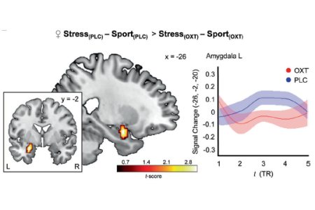

Do you know the smell of fear? Did you know that anxious individuals exhibit heightened sensitivity towards stress signals that they smell in sweat? But can oxytocin, a hormone that is associated with social affiliation, diminish behavioral and neural responses to social chemosensory stress cues? To provide an answer to this question, social neuroscientists from Bonn and biopsychologists from Bochum tested subjects in a forced-choice emotional face recognition task with neutral to fearful stimuli while they were exposed to both sweat stimuli and control odors following intranasal oxytocin or placebo (PLC) administration, respectively. Beforehand, axillary sweat had been obtained from healthy male donors undergoing the Trier Social Stress Test (stress) and bicycle ergometer training (sport). On the neural level, oxytocin reduced stress-evoked responses in the amygdala. It also reinstated the functional connectivity between the anterior cingulate cortex and the fusiform face area that was disrupted by stress odors under placebo. These findings open the door to a completely new role for oxytocin in the modulation of chemosensory stress communication. Mechanistically, this effect appears to be rooted in a downregulation of stress-induced limbic activations and concomitant strengthening of top-down control descending from the anterior cingulate cortex to the fusiform face area.
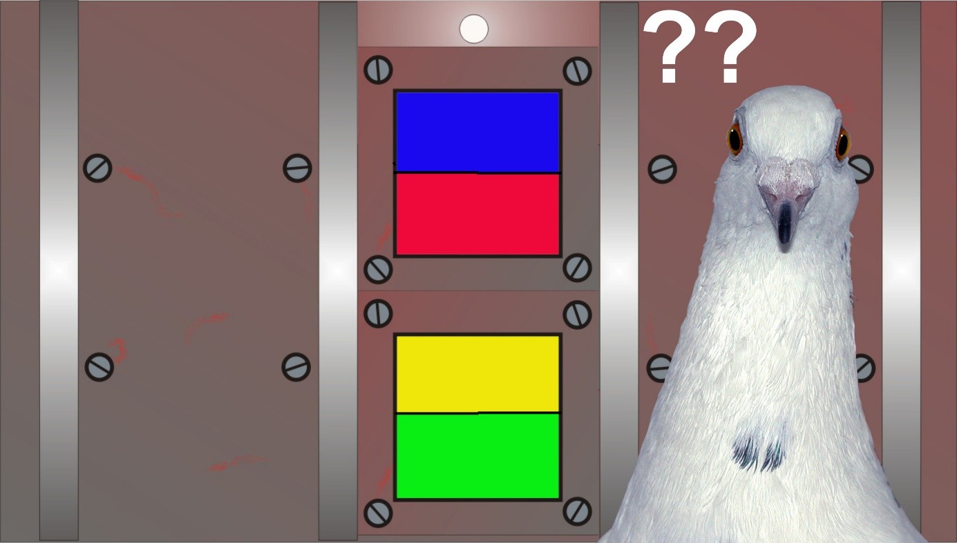

Meta-control describes the phenomenon that one hemisphere takes charge of the response when the two hemispheres plan different actions. The neural fundament of meta-control is possibly a sort of power-play across the commissures. To test this assumption, biopsychologists from Bochum and neurophysiologists from the Chinese Academy of Sciences (Beijing) tested pigeons in a task, in which each hemisphere aimed to peck on a different color pattern. In the critical trials, the animals had to choose between pecking keys that elicit different responses from each hemisphere. The experimental pigeons were tested in this meta-control task both before and after transection of the commissura anterior. This fiber pathway is the largest pallial commissure of the avian brain. The results revealed that meta-control seemed indeed to be modified by interhemispheric transmission via this commissural system.
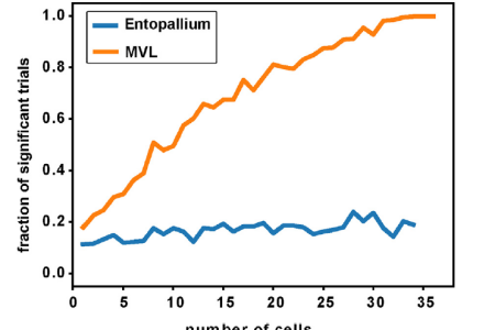

The world surrounding us offers an endless variety of scenes and objects. But just by looking around we effortlessly categorize them in categories like furniture, people, etc. in a blink of an eye. How is this possible? To approach this question, biopsychologists and theoretical neuroscientists from Bochum confronted pigeons with a large number of photographs of objects (vegetables, animals, household items, human faces, etc.), while recording from neurons at different hierarchies of their visual system. Using a linear classifier, the scientists found that the population activity in the primary visual entopallium did not code for any category within these pictures. The higher visual associative area MVL, however, distinguished easily between animate and inanimate objects. Even just about 30 MVL-neurons were sufficient to disambiguate between these categories. It is important to note that such a categorization was not required by the pigeons but emerged by itself from the population code. It seems that visual stimuli are processed along the visual pathways in a hierarchical manner. With every stage in the hierarchy, neurons respond selectively to more complex features, transforming the population representation of the stimuli and making it easy to read-out category information in higher visual areas.
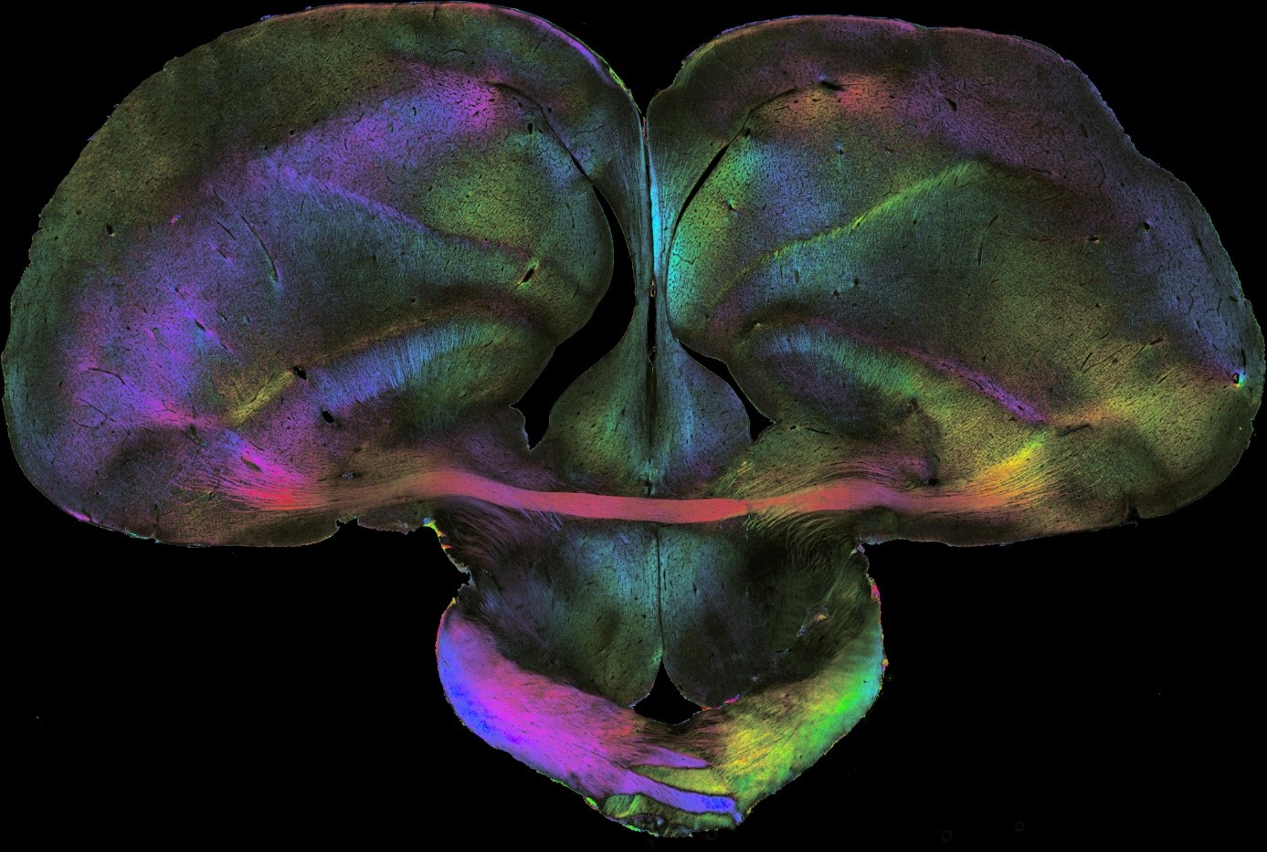

Functional brain asymmetries depend both on hemisphere-specific factors and lateralized commissural interactions. But how are these factors organized in the brain and how are they translated into behavior? Because birds are visually lateralized, biopsychologists from Bochum and neurophysiologists from the Chinese Academy of Sciences (Beijing) tested pigeons monocularly in a color discrimination task while recording from single visuomotor forebrain neuron. All birds learned faster and responded quickly with the right eye and left hemisphere. This asymmetry depended on several factors. One of them concerns the interhemispheric interaction via the commissura anterior: The dominant left hemisphere was able to adjust the timing of activity patterns of right hemispheric neurons via asymmetrical commissural interactions, such that the right hemisphere came too late to control the response. These results imply that hemispheric dominance in birds is realized in part by shifts of the contralateral spike time. Thus, this paper sketches a completely novel picture on the mechanisms of interhemispheric interactions and suggest that the control of the action time of the other hemisphere is a key variable for brain asymmetries. Organisms with lateralized brains need a commissural system that enables the two hemispheres to flexibly switch back and force between recruiting contralateral resources or inhibiting them. According to the results of this paper, this is realized in pigeons by adjusting contralateral spike times. If a similar mechanism could be discovered in humans, it could solve a core riddle of brain asymmetries in our species.


Juvenile idiopathic arthritis (JIA) is a chronic inflammatory disease that starts before the age of 16 and is associated with pain and swellings of the joints but often also with severe fatigue. Especially this condition is characterized as overwhelming and as a serious reduction of life quality. Understanding the complex interactions between the immune and the neural systems of these patients could provide novel possibilities for Pharma-Food interventions in order to improve the quality of life of patients suffering from chronic inflammation. To this end, pharmacologists and rheumatologists from Utrecht university as well as biopsychologists from Bochum analyzed serum samples from JIA patients with an extremely sensitive high-performance liquid chromatography and discovered signatures of an important imbalance between glutamate and various monoamines. These major changes of the brain’s neurochemistry could explain the negative implications of JIA for well-being, including fatigue, cognition, anxiety, and depression.


Do animals prefer to earn their food by hard work instead of getting it for free? Well, many studies indeed show that rats may prefer earned over free food. This unexpected phenomenon is called “contra-freeloading”. In rats the transmitter dopamine—which is involved in incentive motivation and effort— facilitates the occurrence of this preference. Does this also apply to pigeons? Biopsychologists from Bochum tested this in pigeons of which some received a dopamine D2/3 receptor agonist. The birds were exposed to a bowl that contained grains only (easy food) and a bowl that contained grains covered with sawdust (harder food). The animals were tested under various conditions (hungry vs. satiated, successive vs. simultaneous presentation of the options, different sequences of presentation, different doses of dopamine receptor agonist, etc.). There was a single consistent result: Pigeons preferred easy food over difficultly earned one. The scientists could show that this was not due to the option that their drug intervention did not work: It worked; but, different from rats, its action consisted of reducing—rather than magnifying— the attractiveness of the harder food option. Overall, pigeons seem to enjoy chilling more than investing effort. Sounds familiar, isn’t it?


On Wednesday, the 19th of December 2018, Mehdi successfully defended his PhD thesis entitled "Establishing a Novel fMRI Approach to Investigate Visual Cognitive Properties in Pigeons". Mehdi’s PhD studies are an incredible tour de force through all complexities and difficulties that face a brave person who is establishing a completely novel technical approach to realize a breakthrough in cognitive neuroscience. The committee, consisting of Annemie van der Linden (Univ. Antwerp), Martina Manns, Nikolai Axmacher and Onur Güntürkün, were all deeply impressed by these achievements but nevertheless grilled Mehdi heavily by dozens of intricate questions. Mehdi stood the ground until everybody was convinced that he fully deserves to be awarded a PhD. Accompanied by a long applause of all Biopsycho’s, he then put on his mortar board that incorporated a small MRI system to scan in the future even smaller animals. Congratulations Mehdi! We all are very proud of you!

The phrase ‘two eyes with wings’ from Rochon-Duvigneaud (1943) most vividly describes pigeon’s visual abilities and their visual dependence in perceiving the environment. The nidopallium frontolaterale (NFL) is one of the most important visual structures in the pigeon brain and converges information from the avian tectofugal and thalamofugal pathways. It is therefore interesting to see whether this visual structure is involved in extinction learning, the fundamental learning mechanism in adaptation to the changing situations. We recruited a within-subject appetitive extinction design, and found out that transient inactivation of the NFL during extinction did not induce any perceptual impairment, but the extinction acquisition was mildly deteriorated. It implies that the encoding of extinction memory entailed the activation of NFL. In addition, rendering inactivation in the NFL during extinction results in an abolishment of the renewal effect, which indicates that NFL is essential in context encoding, possibly in the process of forming specific context-CS-configurations. Hopefully, this species-specific role of NFL in extinction learning may spark new interests in revisiting the mammalian visual cortices in the process of extinction learning.
Gao, M., Lengersdorf, D., Stüttgen, M. C., & Güntürkün, O. (2019). Transient inactivation of the visual-associative nidopallium frontolaterale (NFL) impairs extinction learning and context encoding in pigeons. Neurobiology of Learning and Memory, 158(October 2018), 50–59. https://doi.org/10.1016/j.nlm.2019.01.012

In the 1860s Pierre Paul Broca proclaimed to have found the seat of articulatory language. One hundred and fifty years later, it is unlikely to find a person studying the brain who has not heard of “Broca’s area” – a term designating to a cortical region in the left hemisphere’s frontal lobe. An abundant amount of studies support Broca’s area’s involvement in language processing. However, modern neuroscience has long shifted its central paradigm. No longer do we believe that a single brain region is the sole basis of cognitive function as complex as language, with its numerous sub-functions. The present article discusses the current and histological implications of Broca’s work. Though published as a scientific article, we tried to bridge the gap between review article and a respectful remark. Although we surely “speak with our left hemisphere”, the truth is certainly more complex.
Friedrich P, Anderson C, Schmitz J, Schlüter C, Lor S, Stacho M, Ströckens F, Grimshaw G & Ocklenburg S. (2019). Fundamental or forgotten? Is Pierre Paul Broca still relevant in modern neuroscience? Laterality: Asymmetries of Body, Brain and Cognition, 24:2, 125-138, DOI: 10.1080/1357650X.2018.1489827.


On Wednesday, the 25th of July 2018, Judith succesfully defended her PhD thesis entitled "The ontogenesis of asymmetry in humans – a genetic and epigenetic perspective". Judith impressed the comitee and other visitors of her Defense with a strong, theory-driven presentation of the papers on the genetics and epigenetics of hemispheric asymmetries she had published in her Phd. In true pioneer fashion, she was one of the first researchers to establish epigentic research in hemispheric asymmetries in humans. During the subsequent discussion, Judith was able to answer even the most difficult questions with ease. Due to the excellence of both her written thesis and her thesis defense , the comitee awarded her the rarely given "Summa cum laude" - the highest honor for a PhD thesis in Germany. Congratulations Judith! Your supervisors and the rest of the lab are very proud of you!
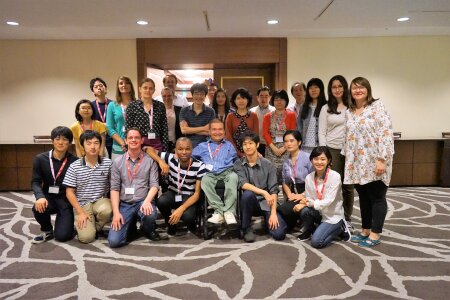

During the last week, the "Brain and Evolution 2018” workshop took place in one of the most exciting cities in Japan: Sapporo. Supported by the DAAD and the Japanese Society for Zoology, students, young academics and senior scientist from the biopsychology lab and different Japanese labs had the opportunity to discuss their projects, plan new experiments and intensify Japanese-German cooperation in the field of comparative neuroscience and brain evolution. From cetacean neuroanatomy to chicken behavior and fish brains (and many other research areas) the summer school covered a wide range of fascinating topics within the field of comparative neuroscience and brain evolution. Despite unfortunately being cut short by an earthquake that hit Sapporo, the summer school was a great experience and received overwhelming positive feedback from all participants. We are looking forward to further cooperations and activities with our old and new friends from Japan!

No matter whether it is in sports or academics, we need to overcome hurdles to reach our goals. In this context, we often ask ourselves why some people get along more successfully than others and where the differences are between those who stumble and those who do not. Individual differences in the ability to initiate self- and emotional-control mechanisms have been related to personal and professional success repeatedly. These differences have been explicitly described in Kuhl’s action-control theory. Although interindividual differences in action control make a significant contribution to our everyday life, their neural foundation remains unknown. To gain deeper insight into the neuronal basics of action control, biopsychologists from Bochum measured action control in a sample of 264 healthy adults and related interindividual differences in action control to variations in brain structure and resting-state connectivity. Their results demonstrate a significant negative correlation between decision-related action orientation (AOD) and amygdala volume. Thus, individuals with larger amygdala volume were less successful to use self- and emotional control mechanisms to achieve a particular goal and in turn, more prone to procrastination and hesitation. Further, the functional resting-state connectivity between the amygdala and the dorsal anterior cingulate cortex (dACC) was significantly associated with AOD. Here less functional connectivity was associated with lower AOD scores. Altered resting-state connectivity between these two brain areas might affect the top-down regulation between the dACC and the amygdala, that is crucial for successful action control. These findings are the first to show that interindividual differences in action control, namely AOD, are based on the anatomical architecture and functional network of the amygdala.

We understand language primarily due to our left hemisphere, which is dominant for linguistic processes. The asymmetry between the left and right hemispheres is easily demonstrated during so-called dichotic listening: When different syllables are presented to either ear, such as "pa" to the left ear and "ba" to the right ear, most people report that they heard the "ba". That is because right ear possesses a more direct pathway to the left, language dominant hemisphere. The effect is also reflected in a speed difference between the left and right hemisphere: Measuring the EEG during dichotic listening reveals faster processing in the left compared to the right hemisphere. But why is the left hemisphere faster? The answer to this question was often believed to lie within a brain region called "Planum temporale", which is bigger on the left than the right hemisphere. Importantly, the left Planum temporale possesses a higher number of neural connections than the right on. This microstructural asymmetry might solve the mystery about the left hemispheric speed advantage. Hence, we tested if there is a direct relation between the processing speed and microstructure in a large sample of almost 100 human participants. Using advanced imaging methods, we measured the density of neural connections of the Planum temporale. Additionally, the processing speed was measured via EEG during dichotic listening. Our results showed that participants with a high density of neural connections in the left planum temporale indeed showed faster processing speed. This finding unreveals a key mechanism for the question why the left hemisphere is so important for language: The high number of neural connections in the left Planum temporale possibly enables a higher precision in the communication between neurons, which in turn leads to faster processing. This superiority in microstructure might be a crucial part of why the left hemispheric became specialized for language.


The MORC1 gene has been suggested as a link between early life stress and major depression in humans as well as in animal models. Here, researchers from the Biopsychology department and the division of Experimental and Molecular Psychiatry investigated DNA methylation in the MORC1 promoter region in healthy human adults. In a non-clinical population, DNA methylation in the MORC1 promoter region was significantly correlated with the Beck Depression Inventory score. In contrast, DNA methylation was negatively associated with birth complications, an indicator of early life stress. These findings further confirm that MORC1 is a stress-sensitive gene and a possible biomarker for depression.
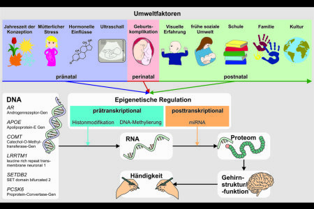

Based on latest research output, researchers from the biopsychology lab were invited to contribute a review article to the prestigious journal BIOspektrum, a molecular biologically oriented journal by the German genetics association. Reviewing the current state of research on handedness, the most prominent of functional hemispheric asymmetries, this German article describes research on genetic and epigenetic factors potentially contributing to handedness ontogenesis. Initially thought to be determined by a single gene, molecular genetic studies have shown that handedness is influenced by multiple genes. However, genetic factors can only explain a quarter of the variance in handedness data. Recent research indicates that epigenetic regulation induces asymmetrical gene expression in the embryonic spinal cord, potentially leading to motor asymmetries. An integration of multiple genetic, epigenetic, and environmental factors is essential to fully comprehend handedness ontogenesis.
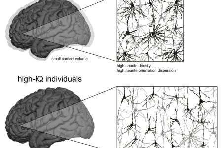

Individuals differ with regard to their cognitive abilities. For more than a century, scientists have been occupied with the question whether such differences in intelligence are associated with differences in the biological properties of the brain. Evidence from modern neuroscience shows that individuals with bigger brains tend to perform better on intelligence tests compared to individuals with smaller brains. However, research also indicates that the brains of intelligent individuals, despite having a comparably larger number of neurons, exhibit less neuronal activity while working on an intelligence test compared to brains of less intelligent individuals. So far, the cortical microstructure underlying these seemingly contradictive findings remained unknown. Here, a group of researchers from the Biopsychology Department collaborated with scientists from Bochum, Berlin and Albuquerque. They administered a matrix-reasoning test to measure intelligence and utilized a special form of in vivo magnetic resonance imaging to assess the amount of dendritic tissue in the cortex. In doing so, it could be shown for the first time that intelligence is inversely related to the number of dendrites connecting cortical neurons. The respective results could be confirmed by a large independent data set from the Human Connectome Project. According to these findings, intelligent brains are characterized by a slim but efficient circuitry between its cortical neurons, enabling high cognitive performance with neuronal activity remaining as low as possible.
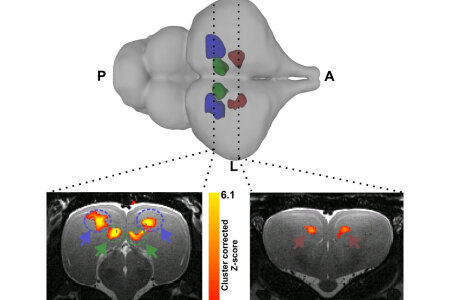

The phylogenetic position of crocodilians in relation to birds and mammals make them an interesting animal model to investigate the evolution of sensory systems in vertebrates. An appropriate, non-invasive method that can be utilized in such studies is fMRI. An international group of researchers from the Biopsychology Department and the School of Anatomical Science at the University of the Witwatersrand (South Africa) now employed fMRI, never previously tested in poikilotherms, to investigate crocodilian telencephalic sensory processing. Juvenile Crocodylus niloticus were placed in a 7T MRI scanner and BOLD signal changes were recorded during the presentation of visual (flickering light at 2-8 Hz) and auditory (simple: chords centered around 1000 or 3000 Hz, and complex: classic music) stimuli. Our findings confirm that the majority of activated regions to both visual and auditory stimuli parallel what has been described for mammalian and avian sensory pathways, indicating conserved sensory processing principles among amniotes. The application of fMRI to ectothermic vertebrates provides an avenue for the application of this method to future functionally related brain research in a broader spectrum of vertebrate species.
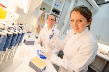

For some decades, single gene explanations have been the most popular models for the ontogenesis of handedness. However, molecular genetic studies revealed only few specific genes exerting small influences on the phenotype. Moreover, non-genetic factors like birth complications and maternal health problems during pregnancy have often been associated with an increased probability of left-handed offspring. Recent research indicates that asymmetric DNA methylation and gene expression in the human fetal CNS contribute to the development of hemispheric asymmetries. Here, a group of researchers from the Biopsychology and the Genetic Psychology Department analyzed DNA methylation in promoter regions of candidate genes for handedness in adult left- and right-handers to investigate whether epigenetic biomarkers of handedness can be identified in non-neuronal tissue. We found that DNA methylation patterns in genes asymmetrically expressed in the fetal CNS predicted handedness direction. Moreover, birth stress was correlated with DNA methylation in NEUROD6, a gene that is asymmetrically expressed in fetal brains. We propose that an integration of genes and environment is essential to fully comprehend the ontogenesis of handedness.
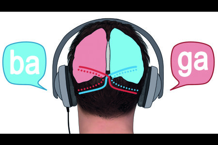

Cognitive control processes play an essential role not only in controlling actions but also in guiding attentional selection processes. In this study, a team of reseachers from Bochum, Bergen and Dresden investigated whether neurobiological mechanisms that affect functional cerebral asymmetries will also modulate cognitive control. Using the Dichotic smartphone app, the research team investigated the association of genetic variation in the handedness-associated gene LRRTM1 and cognitive control in the dichotic listening task. The results show that functional cerebral asymmetries in the language domain are associated with the rs6733871 LRRTM1 polymorphism when cognitive control and top-down attentional mechanisms modulate processes in bottom-up attentional selection processes that are dependent on functional cerebral asymmetries. The results suggest that cognitive control processes are an important factor to consider when being interested in the genetics of hemispheric asymmetries.
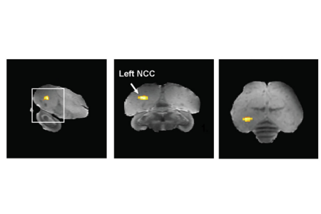

Humans select their partners based on a wide variety of factors; their looks, how much money they have on the bank, or a funny personality. For songbirds, it is all about song. Despite its crucial role in mate choice, little is known about the neurobiological mechanisms underlying the accurate interpretation of courtship song in females. Using a combination of functional resonance imaging (fMRI), immediate early gene expression, and behavioral tests, a group of scientists from Antwerp, Montreal, and Bochum aimed to identify the circuitry involved in the evaluation of mating songs in the zebra finch (Taeniopygia guttata). Females were exposed to either the longer, faster, and more stereotyped courtship song or a neutral song, and this revealed two brain regions, the Mesopallium caudomediale (CMM) and the Nidopallium caudocentrale (NCC), that specifically become active when listening to the mating song. The CMM is a well-known auditory area sensitive to differences in tempo. Since a fast pace is a hallmark of the male courtship song, this was not a very surprising result. More surprising was the activation of the NCC, a ‘hub’ area in the limbic forebrain. This area integrates complex auditory information with sexual imprinting memory of what is desirable of a song. Moreover, the NCC projects to the arcopallium, which is important for coordinating movement. Thus, the NCC is well suited to evaluate the attractiveness of a song and in response coordinate courtship behavior; like calling back to a desirable mate. This study is the first to show the important role of limbic pathways in the evaluation of courtship song and ultimately mate choice of female zebra finches.

On Wednesday, the 14th of February 2018, Patrick succesfully defended his PhD thesis entitled "The Inter- and Intrahemispheric Neural Foundation of Functional Hemispheric Asymmetries". Since the day of his defense fell on Valentine's Day 2018, Patrick showed his love for science by giving the captivated audience a very well received presentation on the triadic model and all the experiments he performed to prove it. After starting in 2014, Patrick has used an impressive methodological array from EEG to fMRI and NODDI imaging to investigate hemispheric asymmetries and interhemispheric interaction and yielded some fascinating insights that he presented in the 25 minute talk. During the subsequent discussion, Patrick was able to answer even the most difficult questions on NODDI imaging and its validation. Obviously, the committee unanimously decided that he had passed. It was decided to award him the prestigous grade of a Dr. rer. nat. with magna cum laude.
Relived, Patrick was presented his PhD hat, inspired by his love for chess and coffee (he has drunken approximately 3500 cups during his PhD).
Congratulations Patrick! Your supervisors and the rest of the lab are proud of you!


On Friday, the 8th of December 2017, Carlotte had her IGSN graduation day. She had already gloriously defended her PhD thesis entitled „Categories in the pigeon brain: From brain function to behavior“ many months before. Even her old Master thesis advisor Miko Colombo had taken all the way from New Zealand for that occasion. But Charlotte is worth it. For sure. In her experimental work she had managed to give the meanwhile long established categorization research in pigeons a completely new direction. Charlotte could demonstrate that we should not look for exemplar or prototype cells in vain but should look for the wisdom of the population code. This provides a completely new way of looking at categorization processes. And she had introduced a novel tool, digital embryos, that is now the trademark of the whole SFB 874. What a success story! The IGSN ceremony was beautiful. Only Onur had his usual bad hair day. But nobody saw that since all eyes were spot on Charlotte.
Congratulations Charlotte! We are proud of you!
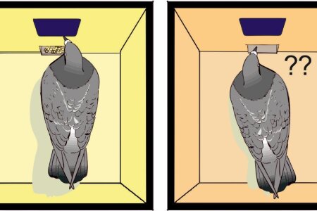

As it is well known to the avian scientific community, a small sector in the avian posterior ventral telencephalon encompasses inextricably intertwined subnuclei which are identified as being of amygdaloid (amygdala) or of somatomotor (arcopallium) nature. Within the SFB 1280, we scrutinized the functional roles of the avian amygdala and the premotor arcopallium in the course of appetitive extinction learning. Since extinction learning is crucially involved in the ability to flexibly acclimatize to the incessantly changing environment which is indispensable for the survival of living organisms, it is of great importance to comprehend the invariant properties of the neural basis of extinction learning. Therefore, we recruited pigeons as our animal model and locally blocked the NMDARs in the avian amygdala and the arcopallium prior to extinction training. We found out that the encoding of extinction memory entailed the activation of amygdaloid NMDARs, while the arcopallial NMDARs were engaged in the consolidation and subsequent retrieval of extinction memory. Furthermore, rendering inactivation in the premotor arcopallium also prompted a general perturbation in the motoric output. The double dissociation between arcopallium and amygdala discerned in the study imparts new insights on the two key components of the avian extinction network. Importantly, the resemblance of our results to the data procured from mammals indicates a shared neural mechanism underlying extinction learning moulded by evolution.
Gao, M., Lengersdorf, D., Stüttgen, M. C., & Güntürkün, O. (2018). NMDA receptors in the avian amygdala and the premotor arcopallium mediate distinct aspects of appetitive extinction learning. Behavioural Brain Research, 343(January), 71–82. https://doi.org/10.1016/j.bbr.2018.01.026

It started with occasional discussions during coffee (“one should finally write a book like that”) and ended, years later, with finally holding this book in hand. The textbook “The Lateralized Brain: The Neuroscience and Evolution of Hemispheric Asymmetries” by Sebastian Ocklenburg & Onur Güntürkün achieves many functions at the same time: It is an entertaining overview of twelve different areas of research on brain asymmetries; each chapter starting with a short story and proceeding with wit and colorful pictures. The book is also a resource for scientists who would like to find a timely overview on different aspects of lateralization with hundreds of references. Last but not least, this book tries to achieve a change of mind in the area of brain asymmetry research. For too long, this field saw itself outside of neurobiology, outside of the animal kingdom, and outside of a serious evolutionary scope. By embedding asymmetry research in these areas, this book works for a kind of science on left-right differences that thrieves for mechanistic explanations of open questions. And in the very end, it was also fun to write it; Sort of.
Ocklenburg, S. and Güntürkün, O., The Lateralized Brain: The Neuroscience and Evolution of Hemispheric Asymmetries, London: Academic Press, 2017.
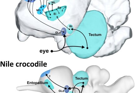

Reptiles and birds are a fascinating group of animals that is most critical to understanding the evolution of vertebrate brains. Birds, who are in fact living dinosaurs, are the only class of vertebrates that can rival mammals with respect to their cognitive abilities. And they do so with brains that are vastly different from ours. This book chapter that was written by biopsychologists from Bochum reviews what we know about reptilian and avian brains in terms of quantitative analyses, structures, and systems. Brains evolved to produce behavior. Therefore, all anatomical and physiological information in this chapter is embedded into a functional framework and provides a rich summary on the current knowledge on archosaur brains.
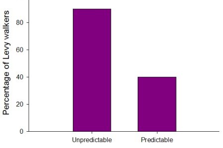

Lévy walks are a property of random movements often observed among foraging animals (and humans), and they might confer some advantages for survival in an unpredictable environment, in comparison with Brownian walks. In animals with a nervous system, specific neurotransmitters associated with some psychological states could play a crucial role in controlling the occurrence of Lévy walks. We argue that incentive motivation, a dopamine-dependent process that in vertebrates makes rewards and their predictive conditioned stimuli attractive, has behavioral effects that may favor their occurrence: incentive motivation is higher when food is unpredictable and it strongly underpins foraging activity. An individual-based computer model is used to determine whether changes in incentive motivation can influence the probability that Lévy walks occur among foraging agents. Our results suggest that they are produced more often under an unpredictable than a predictable food access, and more often in strongly rather than weakly motivated foragers exposed to an unpredictable food access. Also, our motivational framework indicates that the occurrence of Lévy walks are correlated with, but not causally linked to, the number of food items consumed and the ability to store fat reserves. We conclude that Lévy walks can confer some advantages for survival in an unpredictable environment, provided that they appear in foragers with a high motivation to seek food.
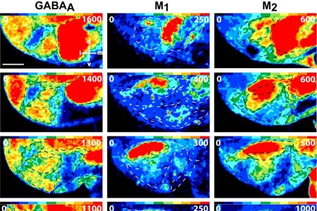

The avian arcopallium/amygdala complex is the abyss of neuroanatomists. You think that the mammalian amygdala is complex? Then come and see the bird version. Here, a bewildering number of limbic (amygdala) and premotor (arcopallium) subnuclei are interwoven on smallest conceivable space. Neuroanatomists from Düsseldorf and biopsychologists from Bochum now took the challenge to map this area by a painstaking quantitative analysis of 12 different transmitter receptor binding sites, combined with a detailed analysis of the cyto- and myelo-architecture. Their approach not only revealed newly discovered subregions but also resulted in a novel map of this most complex area of the bird pallium and striatum. After having accomplished this, the scientists compare the receptor architecture of the subregions to their possible mammalian counterparts and come to a novel interpretation of many of the scrutinized subregions.
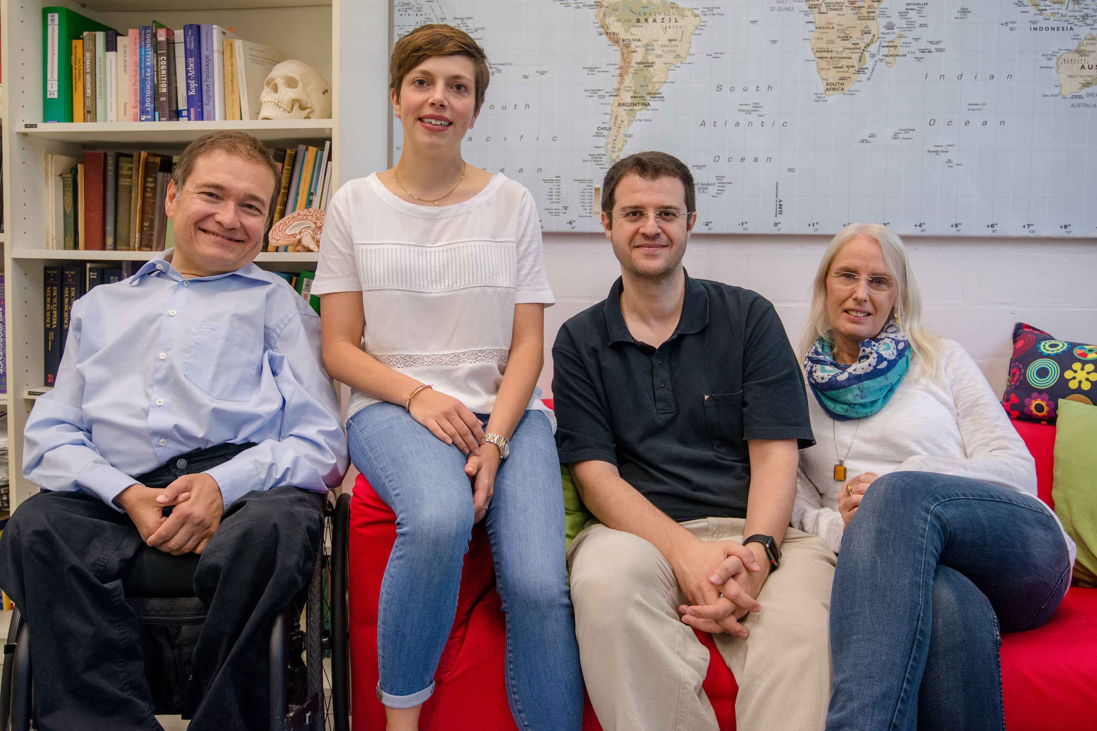

School achievement is highly predictive of future success. Students with better grades achieve better results at university and perform better in job-related contexts and receive higher salaries. Thus, variables predicting school achievement are of interest to teachers, recruiters, and psychologists. Besides cognitive abilities and personality traits, differences in volition are likely to influence the individual’s performance. Biopsychologists from Ruhr University Bochum, therefore, analyzed the influence of volitional abilities in terms of action control on secondary education grading. Their results indicate that action orientation after failure (AOF) and decision-related action orientation (AOD) are associated with secondary school achievement. Furthermore, a multiple regression analysis revealed that AOF and AOD make unique contributions toward predicting final grade, beyond the effects of prominent influencing factors as fluid intelligence, conscientiousness, and sex. Remarkably, the predictive value of conscientiousness did not prove to be unique in nature but was mediated by AOD. The same applies to sex differences in academic achievement. Thus, the influence of sex on final grade was mediated by AOF and AOD. In summary, the study suggests that volition is an essential predictor of achievement in secondary education. Therefore, it's highly recommendable to include measures of volition into future studies investigating the noncognitive correlates of school achievement.
Schlüter, C., Fraenz, C., Pinnow, M., Voelkle, M. C., Güntürkün, O., & Genç, E. (2017, November 16). Volition and Academic Achievement: Interindividual Differences in Action Control Mediate the Effects of Conscientiousness and Sex on Secondary School Grading. Motivation Science.Advance online publication. http://dx.doi.org/10.1037/mot0000083
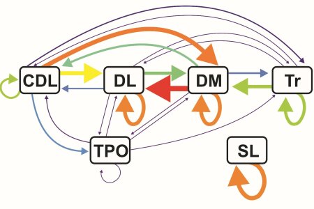

The Hippocampus is a key structure to understand cognition. Unfortunately, we still lack proper knowledge on the interaction between the different hippocampal subregions in birds. To fill this gap, Biopsychologists from Bochum conducted a resting-state fMRI experiment in awake pigeons in a 7-T MR scanner. A voxel-wise regression analysis of blood oxygenation level dependent (BOLD) fluctuations was performed in 6 distinct hippocampal areas, to establish a functional connectivity map of the avian hippocampal network. This first such study in awake birds revealed the functional backbones of information flow within the pigeon hippocampal system. In summary, these findings uncovered a structurally otherwise invisible architecture of the avian hippocampal formation by revealing the dynamic blueprints of this network.
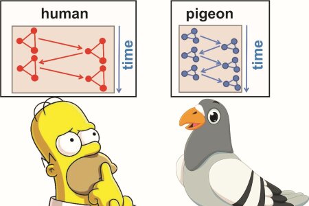

Birds have no cortex and mostly have small brains. How then is it possible that they can master so many complex cognitive tasks? It is known that birds have much more neurons than expected. Therefore, the avian pallium (mostly corresponding to the cortex) is characterized by small, extremely tightly packed neurons. It is conceivable that signal processing could be faster in such a brain as a result of a higher speed of propagation of activation between neighboring assemblies, resulting in faster switch times between neighboring neuronal representations of behavioral goals. Biopsychologists from Bochum and Dresden tested this idea and now indeed report that pigeons are on par with humans when a multitasking experiment demands simultaneous processing resources. However, when task conditions are slightly altered such that high speed of switching between pallial/cortical assemblies is required, pigeons show faster responses than humans. The scientists proposed that this superiority of pigeons could be a consequence of their miniaturized pallium with its high neuronal densities. Inter-neuron distances are on average 1.82 times smaller in pigeons compared to humans, possibly enabling fast activation propagation between neighboring assemblies. This then could represent a key advantage of the non-cortical avian telencephalon over a cortical forebrain.
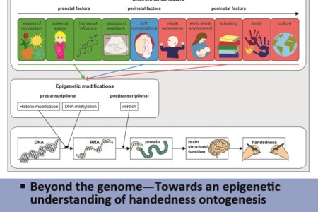

One of the essential organizational principles of invertebrate and vertebrate neurobiology is asymmetric organization of functional or structural properties within the central nervous system. For handedness, the most widely investigated form of hemispheric asymmetries in humans, single gene explanations have been the most popular ontogenetic model in the past. However, while classic family studies suggest heritability of up to 0.66 for handedness, molecular genetic studies revealed only few specific genes contributing to a small amount of phenotypic variance. The divergence between heritability estimates from family studies and results obtained from molecular studies has been referred to as the missing heritability problem, which could partly be accounted for by heritable epigenetic mechanisms. Here, we review recent findings describing non-genetic influences on handedness from conception to childhood. We aim to advance the idea that epigenetic regulation might be the mediating mechanism between environment and phenotype. Recent findings on molecular epigenetic mechanisms indicate that particular asymmetries in DNA methylation might affect asymmetric gene expression in the central nervous system that in turn mediates handedness. We propose that an integration of genes and environment is essential to fully comprehend the ontogenesis of handedness and other hemispheric asymmetries.
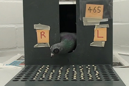

It is still an unanswered question how genetic and environmental factors interact to establish a lateralized brain. In pigeons, the left-hemispheric dominance for visuomotor control depends on asymmetrical light stimulation during embryonic development. In the current study, we explored in how far dominances of the right brain side are determined in parallel. A typical right-hemispheric specialization is visuospatial attention as indicated by enhanced attention to the left visual field. This attentional bias can be demonstrated in a food cancellation task where pigeons have to peck grains regularly scattered on an area in front of them. Comparing the pecking pattern of pigeons with and without embryonic light experience shows that light-exposed pigeons displayed a preference to peck into the left hemifield, supporting a right hemispheric dominance for visuospatial attention. Light-deprived birds on the other hand, displayed an attentional bias to the right halfspace. Thus, light incubation does not simply induce an asymmetry of visuospatial asymmetry; an already present right hemifield bias is rather shifted towards the contralateral left hemifield by asymmetrical light incubation. Moreover, light-exposed pigeons displayed enlarged attentional visual fields extending into the contralateral visual halfspace with each eye compared. These data indicate that asymmetrical embryonic light experience modifies processes regulating biased visuospatial attention and hence, support the impact of light onto the emergence of functional dominances in both brain sides.
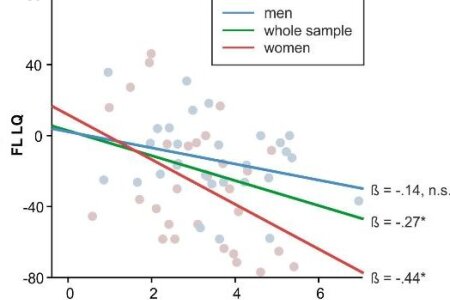

The dichotic listening task is the most widely used paradigm measuring language lateralization. The original non-forced condition consists of different consonant-vowel syllables, i.e. /ba/ and /ga/ presented to the left and right ear, respectively. Most subjects tend to report the syllable presented to the right ear: This so-called right ear advantage reflects left-hemispheric dominance for language. In the forced-attention conditions, subjects are instructed to only attend to input from one ear. Previous studies from the Biopsychology and other labs indicate that heritability is low, which is in line with an epigenetic contribution to language lateralization. The Brandler-Paracchini model of hemispheric asymmetries proposes that genes establishing ciliogenesis and bodily asymmetries affect the development of brain midline structures and language lateralization. KIAA0319 is a promising candidate gene, as it is involved in asymmetrical language processing and ciliogenesis. Here, a group of researchers from the Biopsychology and the Genetic Psychology Department analyzed DNA methylation in the KIAA0319 promoter region to investigate whether epigenetic markers of language lateralization can be identified in non-neuronal tissue. DNA methylation in the KIAA0319 promoter region was predictive for performance in the forced-left and forced-right conditions, but not for performance in the non-forced condition. This is consistent with an effect of DNA methylation on cognitive aspects of language lateralization within the context of the Brandler-Paracchini model of hemispheric asymmetries.
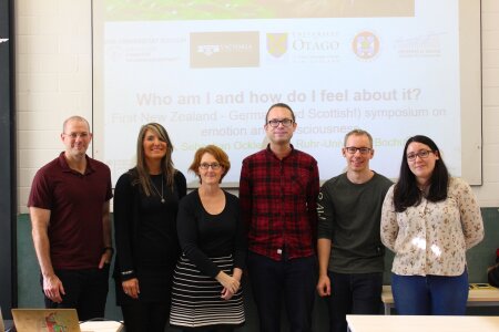

On Friday, 20.10.2017, the first New Zealand - German symposium on emotion and consciousness entitled "Who am I and how do I feel about it?" took place at Ruhr-University Bochum.
Part of an exchange between Victoria University of Wellington and Ruhr-University Bochum funded the initiative for technology and science exchange between New Zealand and Germany, the symposium was a great success.
Hosted by Sebastian Ocklenburg from the biopsychology lab, the symposium had a diverse line-up of speakers from both countries, including Gina Grimshaw from Victoria University of Wellington and Catrona Anderson from the University of Otago, as well as David Carmel from the University of Edinburgh, Jutta Peterburs from Heinrich-Heine-University Düsseldorf and Julian Packheiser from the biopsychology lab. The room was packed with attendants who enjoyey vivid discussion on the nature on emotion and consciousness in both humans and non-human animals.
We are already looking forward to the second symposium!
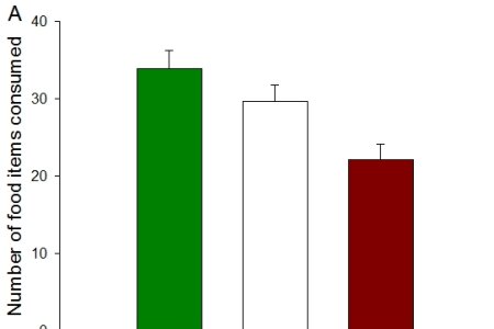

Unpredictable rewards increase the vigor of responses in autoshaping (a Pavlovian conditioning procedure) and are preferred to predictable rewards in free-choice tasks involving fixed- versus variable-delay schedules. The significance those behavioral properties may have in field conditions is currently unknown. However, it is noticeable that when exposed to unpredictable food, small passerines – such as robins, titmice, and starlings – get fatter than when food is abundant. In functional terms, fattening is viewed as an evolutionary strategy acting against the risk of starvation when food is in short supply. But this functional view does not explain the causal mechanisms by which small passerines come to be fatter under food uncertainty. Here, it is suggested that one of these causal mechanisms is that involved in behavioral invigoration and preference for food uncertainty in the laboratory. Based on a psychological theory of motivational changes under food uncertainty, we developed an integrative computational model to test this idea. We show that, for functional (adaptive) reasons, the excitatory property of reward unpredictability can underlie the propensity of wild birds to forage longer and/or more intensively in an unpredictable environment, with the consequence that they can put on more fat reserves.
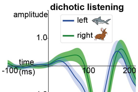

When the two hemispheres receive different sensory input, one might think of a race in which the faster hemisphere dominates our perception.
This can be demonstrated with the so-called “dichotic listening task”. Here, two different vowels are presented simultaneously; one to the left ear that reports to the right hemisphere and one to the right ear, which communicates to the left hemisphere. However, since the left hemisphere is specialized in analyzing vowels, this task is more like a swimming contest between a shark (the left hemisphere) and a rabbit (right hemisphere). Hence, the speed difference between the hemispheres is in favor of the left one.
By using the dichotic listening task in an EEG setting, we measured this hemispheric timing difference. Furthermore, by using MRI techniques, we found that the timing difference is smaller in participants with a better microstructure in the posterior part of the corpus callosum, a white matter commissure that is often called the bridge between the two hemispheres.
Hence, in our metaphor, this bridge seems to help the rabbit across the water, thus equalizing its chances in the swimming competition against the shark.
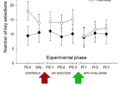

When rodents are given a free choice between a variable option and a constant option, they may prefer variability. This preference is even sometimes increased following repeated administration of a dopamine agonist. The present study was the first to examine preference for variability under the systemic administration of a dopamine agonist, apomorphine (Apo), in birds. Experiment 1 tested the drug-free preference and the propensity to choose of pigeons for a constant over a variable delay. It appeared that they preferred and decided more quickly to peck at the optimal delay option. Experiment 2 assessed the effects of a repeated injection of Apo on delay preference, in comparison with previous control tests within the same individuals. Apo treatment might have decreased the number of pecks at the constant option across the different experimental phases, but failed to induce a preference for the variable option. In Experiment 3, two groups of pigeons (Apo-sensitized and saline) were used in order to avoid inhomogeneity in treatments. They had to choose between a 50% probability option and a 5-s delay option. Conditioned pecking and the propensity to choose were higher in the Apo-sensitized pigeons, but, in each group, the pigeons showed indifference between the two options. This experiment also showed that long-term behavioral sensitization to Apo can occur independently of a conditioning process. These results suggest that Apo sensitization can enhance the attractiveness of conditioned cues, while having no effect on the development of a preference for variable-delay and probabilistic schedules of reinforcement.
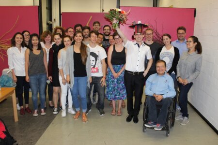

On Friday, the 25th of August 2017, Martin defended his PhD thesis entitled „ The canonical circuit of the avian forebrain “. Martin presented us a fantastic tour de force through the circuitry of the avian pallium and was able to embed his findings and ideas into a detailed framework of previous studies and theories (he concluded that pretty much all of them were (partly) wrong). During the subsequent discussion the word most often used by the reviewers and Martin was “column”. We all are pretty sure that Martin was dreaming of columns the next night. Martin could nicely respond to all questions and successfully defended his ideas. It was just great! Obviously, the committee unanimously decided that he had performed extremely well and decided to award him the rarely awarded grade of a Dr. rer. nat. with summa cum laude. Afterwards, Martin proudly could wear his mortar board that was decorated with… (guess what?): of course columns!!!
Congratulations Martin! We are proud of you!

It is our pleasure to announce the first " First New Zealand - German symposium on emotion and consciousness" which will take place on
Friday, October 20th 2017
12:00 to 15:00
Ruhr-University Bochum, GAFO 05/609
Speakers:
Gina Grimshaw (School of Psychology, Victoria University of Wellington, New Zealand)
David Carmel (Department of Psychology, University of Edinburgh, Scotland)
Jutta Peterburs (Biopsychology, Heinrich-Heine-University Düsseldorf, Germany)
Julian Packheiser (Biopsychology, Ruhr-University Bochum, Germany)
Catrona Anderson(Department of Psychology, University of Otago, New Zealand)
Hosted by:
Sebastian Ocklenburg (Biopsychology, Ruhr-University Bochum, Germany)
All guests are welcome!
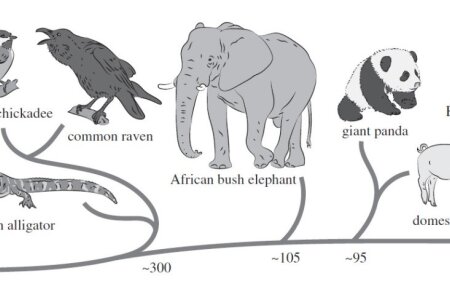

Writing over a century ago, Darwin hypothesized that vocal expression of emotion dates back to our earliest terrestrial ancestors. If this hypothesis is true, we should expect to find cross-species acoustic universals in emotional vocalizations. Previous studies suggest that acoustic attributes of aroused vocalizations are shared across many mammalian species, and that humans can use these attributes to infer emotional content. But do these acoustic attributes extend to non-mammalian vertebrates? In this study, a group of philosophers from Brussels and Bochum, Biopsychologists from Bochum and many animal vocalization specialists from all over the world asked human participants to judge the emotional content of vocalizations of nine vertebrate species representing three different biological classes—Amphibia, Reptilia (non-aves and aves) and Mammalia (see picture to get to know the animals used and to gain insight about their phylogenetic distance to humans in years). They found that humans are able to identify higher levels of arousal in vocalizations across all species. This result was consistent across different language groups (English, German and Mandarin native speakers), suggesting that this ability is biologically rooted in humans. Our findings indicate that humans use acoustic parameters that indicate a shift to higher frequencies to identify higher arousal vocalizations across species. These results suggest that fundamental mechanisms of vocal emotional expression are shared among vertebrates and could represent a homologous signalling system. This study attracted a huge amount of attention from media across the globe and even made it to the news outlet of the Science magazine (http://www.sciencemag.org/news/2017/07/can-you-tell-whether-frog-excited-just-listening-its-voice).

There is good evidence that some bird species can “see” the pole via their right eye and thus with their left brain hemisphere. This lateralization develops during the first winter and initially shows important plasticity. During the first spring migration, magnetic compass asymmetry can even be temporarily abolished by covering the right eye. In the present paper, biologists from Frankfurt and biopsychologists from Bochum used the migratory orientation of robins to analyze the circumstances under which the lateralization of magnetic compass orientation can be altered. It turned out that in young animals already a period of 1½ h in which the dominant right was occluded was sufficient to provide the left eye the ability for successful compass orientation. However, this effect was rather short-lived, as the right eye dominance recurred shortly thereafter again. In addition, training the left eye of robins to adjust to “unnatural” magnetic intensities was possible but was not transferred to the dominant right-eye system. Taken together, the asymmetry of magnetic compass orientation goes through stages of plasticity in the early life of birds. During this time many aspects can be altered and even the dominance direction can be altered, albeit only for short periods. This paper was awarded with the cover of this special issue of the journal “Symmetry”.
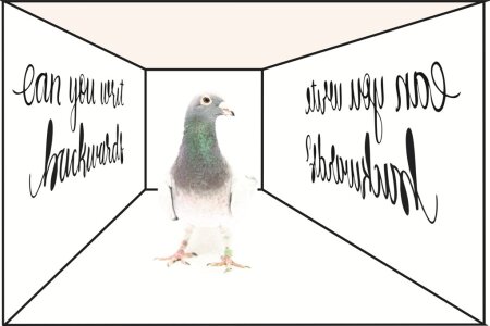

Many children pass through a mirror stage in reading, where they write individual letters or digits in mirror and find it difficult to correctly utilize letters that are mirror images of one another (e.g., b and d). This phenomenon is thought to reflect the fact that the brain does not naturally discriminate left from right. Indeed, it has been argued that reading acquisition in humans involves a learning stage in which this default process is inhibited top-down. In the current study, we tested the ability of literate pigeons, which had learned to discriminate between 30 and 62 words from 7832 nonwords, to discriminate between words and their mirror counterparts. Subjects were sensitive to the left–right orientation of the individual letters, but not the order of letters within a word. This finding may reflect the fact that, in the absence of human-unique top-down processes, the inhibition of mirror generalization may be limited.

Every learning event is embedded in a context, but not always can we subsequently recall this context. Why is context sometimes an integral part of the memory and sometimes not? A good way of studying this is to concentrate on extinction learning. Here, it is assumed that context is not learned for initial acquisition but perfectly learned for the extinction period. While the neuronal mechanisms underlying contextual control of extinction are well known, the mechanisms of (non)learning during acquisition are mostly unknown. Here, we tested the hypothesis that shifting attention to critical cues is less likely during acquisition than during extinction and that this is the reason why context is less learned during acquisition than during extinction. We further assumed that these attentional mechanisms are controlled by activation of glutamatergic NMDA-receptors at prefrontal level. Thus, we antagonized NMDA receptors with AP5 in the pigeon nidopallium caudolaterale (NCL), the functional analogue of mammalian prefrontal cortex, during the concomitant acquisition and extinction of conditioned responding to two different stimuli. Indeed, NMDA receptor blockade resulted in an impairment of extinction learning, but left the acquisition of responses to a novel stimulus unaffected. Critically, when responses were tested in a different context in the retrieval phase, we observed that NMDA receptor blockade led to the abolishment of contextual control over acquisition performance. Thus, learning without functional NMDA receptors in NCL possibly results in a loss of contextual gain on the learned association, possibly via the modulation of attentional mechanisms. To come back to our initial question of why we sometimes learn the context and sometimes not: It depends on how much our attention is switched to the context during learning. If we pay attention, NMDA receptors at prefrontal level play a key role.
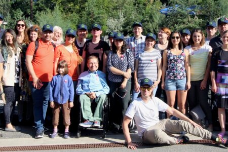

On the 28th of July 2017, the Biopsychology and the Avian Cognitive Neuroscience lab as well as our guests Olga Lazareva & Martin Acerbo (of course incl. Valerie) departed towards the Nuremberg zoo for our yearly retreat. Our friend and colleague Lorenzo von Fersen had invited us to use their facilities for our presentations on the question “What is it that you would like to discover in the next year(s)?” We started in the afternoon with a firework of presentations by diverse groups and individuals. In the evening we jointly visited the traditional restaurant Barfüßer to get accustomed to Bavarian food and kegs of Bavarian beer. On the next morning we had some more talks and then visited the zoo and could participate in an experiment on magnetic perception in dolphins. Towards the late afternoon we all travelled back to Bochum. Everybody deeply enjoyed our stay and we all were convinced that our next retreat should last one day longer.
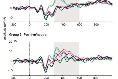

Hemispheric asymmetries are a major organizational principle in human emotion processing, but their interaction with prefrontal control processes is not well understood. In the present study, researchers from the biopsychology lab, the University of Münster and the University of Wellington in New Zealand use a lateralized emotional paradigm to assess cognitive control processes and recorded ERP components. Electrophysiologically, Nogo-P3 responses were greater for emotional than for neutral stimuli, an effect driven primarily by an enhanced response to positive images. Hemispheric asymmetries were also observed, with greater Nogo-P3 following left versus right visual field stimuli. However, the visual field effect did not interact with emotion. We therefore find no evidence that emotion-related asymmetries affect response inhibition processes.
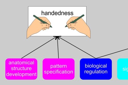

Handedness and language lateralization have often been thought to share a common ontogenetic basis. Although specific genetic influences contributing to each phenotype have been identified, no gene has been found to be involved in both phenotypes. However, it is still possible that genes associated with handedness and genes associated with language lateralization share similar gene functions. Here, a group of researchers from the biopsychology lab conducted gene ontology analyses to identify the amount of overlap in functional gene groups between the phenotypes. Results showed that genes associated with handedness are involved in anatomical structure development, pattern specification (especially asymmetry formation) and biological regulation. Genes associated with language lateralization are involved in responses to different stimuli, nervous system development, transport, signaling, and biological regulation. While “handedness genes” mostly contribute to structural development, genes involved in language lateralization rather contribute to activity dependent cognitive processes. This finding has been confirmed by disease association analysis revealing that genes associated with handedness are involved in with diseases affecting the whole body, while genes associated with language lateralization were specifically involved in mental and neurological diseases. These findings further support the idea that handedness and language lateralization are ontogenetically independent, complex phenotypes.


About 90% of the population is right-handed, which has often been proposed to result from a single gene. However, molecular studies rather support the idea that handedness is determined by a multitude of small, possibly interacting genetic and non-genetic influences. Here, scientists from the biopsychology lab, the genetic psychology lab and several departments of the medical faculty of Dokuz Eylül University in Izmir performed whole exome sequencing in nine left-handed members of a Turkish family with history of consanguinity in order to detect influences of rare mutations on handedness. Quantitative trait analysis revealed that rare variants on 49 loci on 26 genes show significant associations with handedness; however, none was functionally relevant for handedness (i.e. involved in left-right axis development or nervous system development). This interpretation was further supported by gene ontology analysis, as functional gene groups were also unrelated to the brain. Taken together, this study revealed no indication for a gene or mutation that could realistically determine handedness.
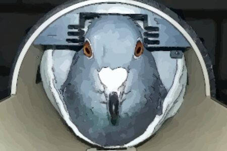

The majority of vertebrate models used in neuroscience are rodents (rats and mice), but increasingly bird species such as zebra finches and pigeons also are used. Whereas zebra finches as songbirds are an excellent model for vocal learning, pigeons typically are used to study mechanisms of learning and memory. However, pigeons also are increasingly tested with cognitive tasks assessing functions, such as sequence acquisition, equivalence learning, categorization, transitive inference, and even orthographic processing. In contrast to rodents, some bird species such as corvids additionally demonstrate high cognitive abilities that are equivalent to those of nonhuman primates. These include complex social interactions, future planning, tool and meta-tool use, mirror self-recognition, and physical problem-solving. To understand the functional and structural organization of the pigeon brain under different conditions, scientists of the Biopsychology Department of the Ruhr-University Bochum developed functional MRI protocols with high temporal resolution. Since transverse (T2) and longitudinal (T1) relaxation times play an important role in optimizing MRI parameters [e.g., contrast level, signal-to-noise ratio], they measured these values for the first time in awake pigeons and rats. The obtained T1 and T2 values for awake pigeons and rats and the optimized habituation protocol will augment future MRI studies with awake animals. The differences in relaxation times observed between species underline the importance of the acquisition of T1/T2 values as reference points for specific experiments.

On May 25, 2017, the DFG Senate Commission for SFBs decided to establish a brand new SFB in Bochum: The SFB1280 Extinction Learning was thus officially established!
This is the first SFB that was born out of our Faculty and the third in Germany given to a Faculty of Psychology. We now have the means to study for four years the behavioral, developmental, neural, and clinical aspects of extinction learning. If successful, the SFB could run for up to 12 years in total. Countless insights, publications, careers, and positions will be born out of that.
The new SFB has 19 project teams. On board are psychologists, medical scientists, theoretical neuroscientists, and biologists from the RUB, and from Essen, Dortmund and Marburg.
more information: RUB news portal / DFG press release
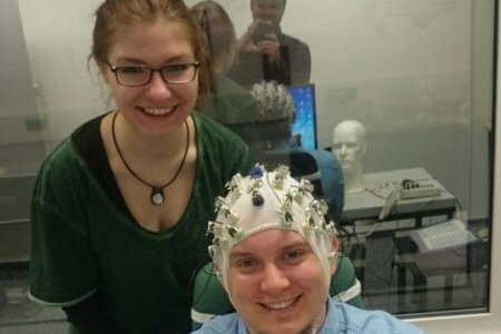

While the two halves of the human brain are connected via white matter commissures, it is difficult to estimate the speed with which the hemispheres share their information.
For a long time, it was believed that a simple behavioral task would do the trick: In one condition, a stimulus is presented on one side while participants should react with the hand of the same side. In the other condition, the stimulus and reacting hand are on different sides. Hence, in the first condition both perception and reaction occur in the same hemisphere, while in the second condition, perception and reaction are processed in different hemispheres. Therefore, the difference in reaction time between the two conditions should reflect the transfer time between the two hemispheres, right? Well, kind of but not quite. For instance, this paradigm is not reliable - If you measure a person today and a week later, the results may vary!
Here, we tested an alternative approach. Instead of relying on reaction times, we calculated the difference in event-related potentials, using the same paradigm with an EEG system. Our results show that with this approach, transfer times measured today are comparable to transfer times measured one year later! Hence, this method allows us to get reliable estimates for the speed of interhemispheric information transfer.

Hundreds of Millions of chicken are raised every year for food consumption. A major problem of crowded poultry farms is severe feather pecking - a detrimental behavior that is also shown by very young chicks. However, not all individuals do this. Why are some birds more aggressive than others? A group of Behavioral Pharmacologists from Utrecht, Veterinary scientists from Wageningen and Biopsychologists from Bochum started to analyze the role of serotonin and dopamine in feather pecking. They had access to two groups of domestic chickens divergently genetically selected for Low Feather Pecking (LFP) and High Feather Pecking (HFP). Indeed, both lines differed both on their serotonin and their dopamine turnover in emotion-regulating and motor areas. Furthermore, HFP-chicks responded more actively in most behavioral tests conducted, and were more impulsive in their way of coping with challenges. Thus, a brain area-specific neurochemical profile may shift personality traits towards more bold but also more aggressive behavior in chicken.
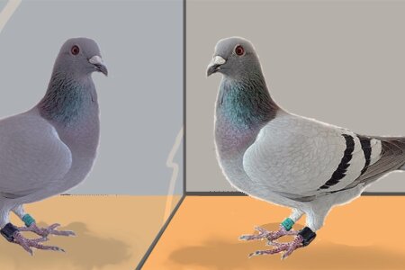

A small number of species are capable of recognizing themselves in the mirror when tested with the mark-and-mirror test. This ability is often seen as evidence of self-recognition and possibly even self-awareness. Strangely, a number of species, for example cats, pigs and dogs, are unable to pass the mark test but can locate rewarding objects by using the reflective properties of a mirror. Thus, these species seem to understand how a visual reflection functions but cannot apply it to their own image. Biopsychologists in Bochum tested this discrepancy in pigeons—a species that does not spontaneously pass the mark test. Indeed, we discovered that pigeons can successfully find a hidden food reward using only the reflection, suggesting that pigeons can also use and potentially understand the reflective properties of mirrors, even in the absence of self-recognition. However, tested under monocular conditions, the pigeons approached and attempted to walk through the mirror rather than approach the physical food, displaying similar behavior to patients with mirror agnosia. These findings clearly show that pigeons do not use the reflection of mirrors to locate reward, but actually see the food peripherally with their near-panoramic vision. A re-evaluation of our current understanding of mirror-mediated behavior might be necessary—especially taking more fully into account species differences in visual field. This study suggests that use of reflections in a mirrored surface as a tool may be less widespread than currently thought.
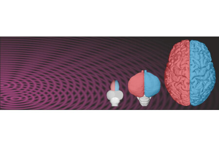

The brains of humans and other animals are asymmetrically organized, but we still know little about the ontogenetic and neural fundaments of lateralizations. Here, we review the current state of understanding about the role of genetic and non-genetic factors for the development of neural and behavioral asymmetries in vertebrates. At the genetic level, the Nodal signaling cascade is of central importance, but several other genetic pathways have been discovered to also shape the lateralized embryonic brain. Studies in humans identified several relevant genes with mostly small effect sizes but also highlight the extreme importance of non- genetic factors for asymmetry development. This is also visible in visual asymmetry in birds, in which genes only affect embryonic body position, while the resulting left-right difference of visual stimulation shapes visual pathways in a lateralized way. These and further studies in zebrafish and humans highlight that the many routes from genes to asymmetries of function run through left-right differences of neural pathways. They constitute the lateralized blueprints of our perception, cognition, and action.
Güntürkün, O. and Ocklenburg, S., Ontogenesis of Lateralization, Neuron, 2017, 94: 249-263.
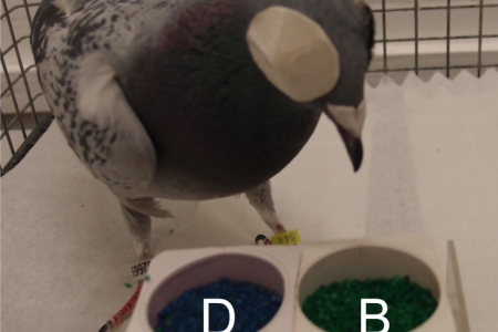

The two hemispheres of the vertebrate brain are characterized by several left-right differences in the analysis and evaluation of information. Such lateralized processing requires intra- and interhemispheric mechanisms mediating exchange, integration, or suppression of information but the underlying functional organization is only basically understood. Researchers from the Biopsychology lab of Bochum therefore explored intrahemispheric integration capacities in pigeons during relational learning. Pigeons were trained in a way that each hemisphere learned only half the information that represented a transitive line (A>B>C>D>E). Subsequently, the hemispheres were tested independently from each other with stimulus pairs that required accessing information from the other brain side. The hemispheres differ in encoding interhemispheric information but both hemispheres were impaired in correct responding when the information was in conflict with directly learnt memory. In sum, the study indicates that interhemispheric communication in pigeons is an active process that integrates intra- and interhemispheric information in a context-dependent and hemispheric-specific manner. Efficient interhemispheric cooperation requires simultaneous activation of both brain sides.


Yes, they left... Annika and Rena were for many years indispensable members of the Biopsychology universe. Hard to believe, but now they left this mother of all labs at the end of February 2017. During their time in the magic lab they made great discoveries. They reconstructed the sequence of the Arc gene, discovered complex pathways to action in the pallium and came extremely close to finally demystifying the critical trigger for visual asymmetry in pigeons. What achievements! What bravery!
Hey Rena and Annika: We will miss you both a lot!!!!

Apes, corvids, and pigeons differ in their pallial/cortical neuron numbers, with apes ranking first and pigeons third. Do cognitive performances rank accordingly? If they would do, cognitive performance could be explained at a mechanistic level by computational capacity provided by neuron numbers. To approach this question, biopsychologists from Bochum and Dunedin (New Zealand) reviewed five areas of cognition (short-term memory, object permanence, abstract numerical competence, orthographic processing, self-recognition) in which apes, corvids, and pigeons have been tested with highly similar procedures. They discovered that in all tests apes and corvids were on par, but also pigeons reached identical achievement levels in three tests. The scientists conclude that higher neuron numbers are poor predictors of absolute cognitive ability, but better predict learning speed and the ability to flexibly transfer rules to novel situations.
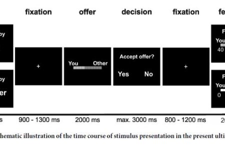

Social context influences social decisions and outcome processing. In the present study a team of researchers from the University of Münster and the Biopsychology in Bochum investigated social context-dependent modulation of behavior and feedback. Adult participants completed an ultimatum game under social observation or not and EEG was recorded. Overall, fewer unfair than fair offers were accepted. Observation decreased acceptance rates for unfair offers. The feedback-locked feedback-related negativity (FRN) was modulated by observation and fairness, with stronger differential coding of unfair/fair under observation. This effect was strongly correlated with individual levels of social anxiety, with higher levels associated with stronger differential fairness coding in the FRN under observation. Behavioral findings support negative reciprocity in the UG, suggesting that social norms overwrite explicit task instructions even in the absence of social interaction. Observation enhances this effect.
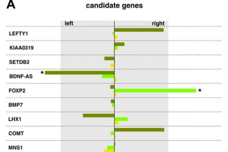

Almost everyone has a preferred hand that excels at most tasks, even though our hands are anatomically almost identical. So far, most researchers assumed that handedness is largely under direct control of variation in certain genes, but the scientific evidence for this assumption has been weak. Epigenetic mechanisms affect gene activity independently from the actual DNA sequence. During human embryonic development, preliminary forms of handedness arise before the motor cortex that controls our movements is connected to the spinal cord that is responsible for movement execution. A multidisciplinary team from Biopsychology and several other departments of the medical and psychological faculties could now show that along with the onset of asymmetric hand movements epigenetic factors (more specifically miRNA and DNA methylation) lead to massive gene expression asymmetries in the spinal cord segments that innervate arms and hands. These findings imply a fundamental shift in our understanding of the ontogenesis of hemispheric asymmetries in humans. For the first time, our study sheds light on the molecular epigenetics of asymmetry formation.
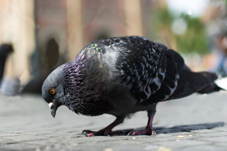

Animals exploit visual information to identify objects, form stimulus-reward associations, and prepare appropriate behavioral responses. The avian “prefrontal” nidopallium caudolaterale (NCL) contains neurons that play a key role in these processes. But do these neurons code for pictorial aspects, stimulus value, or sensorimotor contingencies? To test these questions, biopsychologists from Bochum, Cold Spring Harbor and Mainz subjected pigeons to a Pavlovian sign-tracking paradigm in which visual cues predicted rewards differing in magnitude (large vs. small) and delay to presentation (short vs. long). Subjects’ strength of conditioned responding to visual cues reliably differentiated between predicted reward types and thus indexed valuation. The majority of NCL neurons discriminated between visual cues, with discriminability peaking shortly after stimulus onset and being maintained at lower levels throughout the stimulus presentation period. However, while some cells’ firing rates correlated with reward value, such neurons were not more frequent than expected by chance. Instead, neurons formed discernible clusters which differed in their preferred visual cue. The authors propose that this activity pattern constitutes a prerequisite for using visual information in more complex situations e.g. requiring value based choices.
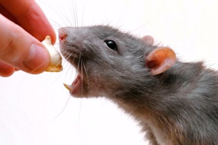

The 5-HT1A/1B-receptor agonist eltoprazine is effective in treating impulsivity disorders, most likely by increasing norepinephrine (NE) and dopamine (DA) levels in the prefrontal cortex. But how eltoprazine affects monoamine release in the medial prefrontal cortex (mPFC), the orbitofrontal cortex (OFC), and the NAc is unknown. It is also unknown whether eltoprazine affects different forms of impulsivity and brain reward mechanisms. Therefore, behavioral pharmacologists and biopsychologists from Utrecht and Bochum investigated the effects of eltoprazine in rats with respect to the activity of the monoaminergic system, the motivation for reward, “waiting” impulsivity in the delay-aversion task, and the “stopping” impulsivity in the stop-signal task. The results clearly showed that eltoprazine increased DA and NE release in both the mPFC and OFC, but only increased DA concentration in the NAc. In contrast, eltoprazine decreased 5-HT release in the mPFC and NAc. Remarkably, eltoprazine decreased impulsive choice, but increased impulsive action. These results further support the long-standing hypothesis that “waiting” and “stopping” impulsivity are regulated by distinct neural circuits, because 5-HT1A/1B-receptor activation decreases impulsive choice, but increases impulsive action.
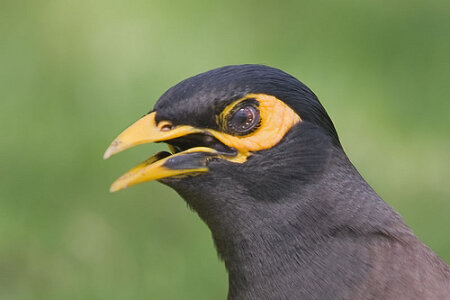

Within a few hundred years, humans have changed vast natural habitats into cities. How do non-human animals cope with these changes? We know that some species disappear, while others exploit the benefits of this new ecology. Possibly, changes induced by urbanized environments cause a novel selection pressure on cognitive abilities. Scientists from Newcastle (Australia), Vienna, and Bochum studied this hypothesis in urban and rural common mynas using a standard visual discrimination task followed by a reversal learning phase. They not only compared the speed of learning but also how quickly each bird progressed through different stages of learning of reversal learning. Based on their reliance on very different food resources, they expected urban mynas to learn and reversal learn more quickly. When quantified from first presentation to criterion achievement, however, urban mynas took more 20-trial blocks to learn the initial discrimination, as well as the reversed contingency, than rural mynas. More detailed analyses at the level of stage revealed that this was because urban mynas explored the novel cue-outcome contingencies for longer, and despite transitioning faster through subsequent acquisition, remained overall slower than rural females. These findings draw attention to fine adjustments in learning strategies in response to urbanization and caution against interpreting the speed to learn a task too easily as a reflection of cognitive ability.
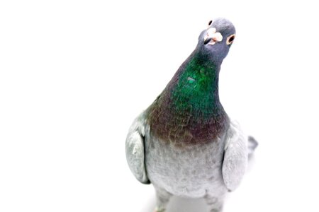

It is widely held that the extinction of a conditioned response is more context specific than its initial acquisition. One proposed explanation is that context serves to disambiguate the meaning of a stimulus. Using a procedure that equated the learning histories of the contexts, Biopsychologists from Cold Spring Harbor, Mainz, Marburg, and Bochum showed in pigeons that the memory of an appetitive Pavlovian association can be highly context specific despite being unambiguous. This result is inconsistent with predictions of the Rescorla–Wagner model of learning but in line with configural accounts of contextual control of behavior. The authors propose an explanatory model in which context serves to modulate the gain of associative strength and which expands upon the configural idea of unitary representations of context and conditioned stimuli.
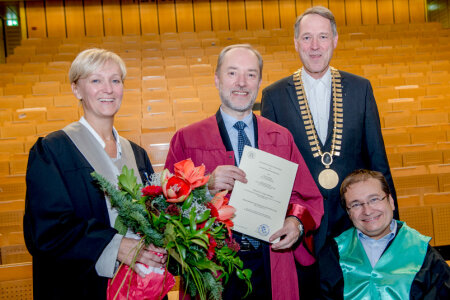

On the 1st of December 2016, The Faculty of Psychology and thus the Ruhr-University Bochum awarded Prof. Dr. Giorgio Vallortigara from Trento University with the title of a Doctor rerum naturalium honoris causa (Dr. rer. nat. h. c.). This is a high and rare award, only given to very distinguished scholars. Within the 51 years of existence of our faculty, this award was before only given twice. Giorgio Vallortigara was awarded for his landmark contributions in comparative cognitive behavioral sciences. Among other insights, he could demonstrate that cognitive abilities that we thought were unique to humans are widespread within animals. He also showed that brain asymmetries are not a privilege of the human species, and that young animals are born with a rich knowledge about the world they are going to live in. Giorgio Vallortigara has published close to three hundred peer-reviewed scientific publications with some of them having appeared in top journals like Nature, Science, PNAS, Plos Biology, or Current Biology. But Giorgio Vallortigara is also a very successful organizer of academic systems. He was instrumental in the creation and foundation of the Center for Mind and Brain Sciences at the University of Trento in Northern Italy. This center is a unique place with a vast technical infrastructure for cognitive neuroscience and behavioral analyses. From the very beginning on, Giorgio Vallortigara was a central figure of this center and became its director from 2002 to 2015.
Congratulations Giorgio!!!


The winter term meeting of the DFG Senate Commission on Graduate Schools had great news for Bochum and Osnabrück: A new Graduate School on Situated Cognition will be established for up to nine years on the Philosophy and the Neuroscience of Mind. Both Philosophers and Biopsychologists of both universities had worked relentlessly on the application. In its very core, the theoretical thrust of the application was to develop an alternative to the outmoded sandwich model of cognition. This sandwich model is the relict of a Fodor’ian thinking about modules. It posits that the mind consists of three main entities; perception, cognition, action. Each of these modules is encapsulated and can be disambiguated from each other. We meanwhile know, however, that cognition encompasses the full extent of information processing that runs from the first aspects of perception till the last step of action generation. Within this framework, emotion, social integration, and decision making will be analyzed using, among others, the concept of embodiment, according to which many cognitive features are shaped by aspects of the body beyond the brain. The picture associated with this message shows such a situation: Forcing us to display the emotion of smiling indeed increases our happiness. Our body shapes our mind. The School has also one PhD-slot for Biopsychologists from Bochum and will start in fall/summer 2017.
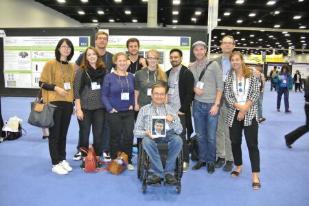

With more than 28.000 researchers in attendence, the Meeting of the Society for Neuroscience (SFN) is the largest gathering of neuroscientists anywhere in the world. This year, it took place at the San Diego Conference Center in California and the Biopsychology lab was well represented with 12 poster presentations, covering diverse topics ranging from optogenetics in pigeons to the epigentics of handedness development in humans. The 4 hour poster presentations were well received by the worldwide neuroscientific community and lead to many fruitful discussions about the cutting edge research performed in the Biopsychology lab.
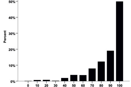

90% of the population is right-handed, but the development of this trait is not well understood. Here, a team of researchers from the biopsychology lab and the human genetics lab at the Faculty of Medicine investigated the role of genes contributing to the differentiation of the left-right axis during embryogenesis for handedness development. The team identified a haplotype on SETDB2 for which homozygous individuals showed a significantly lower lateralization quotient for handedness than the rest of the cohort after correction for multiple comparisons. Moreover, direction of handedness was significantly associated with genetic variation in this haplotype. This effect was mainly, but not exclusively, driven by the sequence variation rs4942830, as individuals homozygous for the A allele of this single nucleotide polymorphism had a significantly lower lateralization quotient than individuals with at least one T allele. These findings further confirm a role of genetic pathways relevant for structural left-right axis differentiation for functional lateralization. Moreover, as the protein encoded by SETDB2 regulates gene expression epigenetically by histone H3 methylation, our findings highlight the importance of investigating the role of epigenetic modulations of gene expression in relation to handedness.

Traditionally, research on language lateralization is conducted in psychological labs, but recently the use of smartphones as test devices is on the rise. Here, a team of researchers from the Biopsychology lab and the Bergen fMRI group used a Apple iOS app to investigate the heritability of language lateralization assessed with the dichotic listening task, as well as the heritability of cognitive control processes modulating performance in this task. Overall, 103 families consisting of both parents and offspring were tested with the non-forced, as well as the forced-right and forced-left condition of the forced attention dichotic listening task, implemented in the iDichotic smartphone app, developed at the University of Bergen, Norway. The results indicate that the typical right ear advantage in the dichotic listening task shows weak and non-significant heritability. In contrast, cognitive factors, like attention focus and cognitive control showed stronger and significant heritability. These findings indicate a variable dependence of different aspects of a cognitive function on heritability and implicate a major contribution of non-genetic influences to individual language lateralization.
This study was also highlighted on the RUB news page!
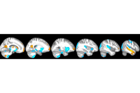

Handedness is thought to originate in the brain, but identifying its structural correlates in the cortex has yielded surprisingly incoherent results. One idea proclaimed by several authors is that structural grey matter asymmetries might underlie handedness.
In the present study, a team from the Biopsychology lab investigated this idea using a new voxel-based morphometry toolbox. While there were several significant left-right asymmetries in the overall sample, no difference between left- and right-handers reached significance after correction for multiple comparisons. These findings indicate that the structural brain correlates of handedness are unlikely to be rooted in macroscopic grey matter area differences that can be assessed with VBM. Future studies should focus on other potential structural correlates of handedness, e.g. structural white matter asymmetries.

According to a new study published by biopsychologists from Bochum and the University of Otago (New Zealand), pigeons are able to discriminate English words from non-words. And they do this by orthographic rules that are identical to those used by humans. This enormous ability demonstrates that orthographic knowledge is no privilege of humans or primates but can be mastered by an animal with just 2.5 gram of brain. To demonstrate this feat, scientists first taught the birds top peck on words (e.g. DONE) shown on a monitor to obtain food. Pecking on non-words (e.g. DNOE) was not rewarded. Slowly the animals learned more and more English words (depending on the individual between 26 and 58) and discriminated them from non-words (about 8,000). Then came the real test: Pigeons were confronted with entirely new English words and non-words. The animals spontaneously discriminated successfully between the two groups of stimuli. Thus, they had learned what is typical for an English word. But how did they do this? A deeper analysis demonstrated that pigeons used two strategies. One was to use the frequency of bigrams in English. The word DONE has three bigrams: DO, ON, and NE. Their average bigram frequency is much higher than those of DN, NO, and OE in the non-word DNOE. The second strategy was the Levenshtein distance between two words which is the minimum number of single-character edits (i.e. insertions, deletions or substitutions) required to change one word into the other. Also humans utilize these two strategies for these kinds of decisions. The brains of birds and humans are vastly different. If still both species use the same strategy, it is likely that evolutionary selection pressure to identify regularities of input statistics are the relevant force that shaped brains during evolution. Whatever the genetically determined general outline of the brain of an animal was, its internal computations had to have the blueprint to solve these tasks. Within one week after its appearance, this paper was among the top 99.95% of all 6,365,760 scientific publications registered so far with respect to international media attention.

On Wednesday, the 24th of August 2016, Rena defended her PhD thesis entitled „ How visual asymmetry starts in pigeons: Characterizing Melanopsin as a potential inducer “. It was just awesome. Rena had tons of data to show, was able to answer each and every question (even the extremely vague ones) and flabbergasted the reviewers by the breadth, ingenuity, and complexity of her experiments and conclusions. Accordingly, the committee unanimously decided that she had performed extremely well and decided to award her the title of a Dr. rer. nat. with magna cum laude. Afterwards everybody could enjoy a party. On the picture Rena is depicted with the whole lab that had good reasons to celebrate one more great scientist from the Biopsychology.
Congratulations Rena! We are proud of you!

From October 4th to 6th 2016, the interdisciplinary workshop “Rational animals? – Comparing human and animal minds from an interdisciplinary perspective” was held at the Ruhr-University Bochum, organized in a joint venture by the Department of Philosophy II and the Biopsychology department. Philosophers, behavioral researchers and neuroscientists from 9 different countries discussed over the course of three days recent findings on the cognitive abilities of non-human animals and how to interpret these findings in the light of modern neuroscience and philosophy. Over 50 participants joint the workshop, listening to talks, visiting a poster session and taking part in several discussion rounds. Although the question if non-human animals are indeed rational could not be answered thoroughly, the workshop was perceived to be highly successful by both the participants and the organizers. Especially for the biopsychology department the workshop proved to be highly successful since two members of our department won the first (Stephanie Lor) and second price (Mehdi Behroozi) for their scientific posters. Steffi & Mehdi congratulations!
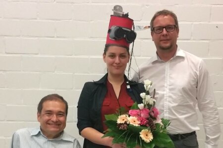

On Friday, the 19th of August 2016, Clara defended her PhD thesis entitled „Stratiales GABA und Handlungskontrolle – Eine kombinierte EEG-, MRS- und fMRI-Untersuchung“. After giving a brilliant presentation, she answered all questions in her typical calm but absolutely clear way. Even the long, rambling and vague questions that were asked by one of the examiners did little to her absolutely solid standing. Accordingly, the committee unanimously decided that she had performed extremely well and decided to award her the title of a Dr. rer. nat. with magna cum laude. Afterwards everybody could enjoy sparkling wine in brilliant sunshine.
Congratulations Clara! We are proud of you!

Throughout life, we learn to associate stimuli with their consequences. But some of the new information that we encounter forces us to abandon what we had previously acquired. This old information is then subject to a new learning process that is called extinction learning that is extremely complex and involves a large number of brain structures. To provide a most recent update on research on extinction learning, Denise Manahan-Vaughan, Onur Güntürkün and Oliver Wolf from the Ruhr-University came together to create an open-access Frontier Research Topic e-book. Their publishing strategy departed from the formulation of three aims: First, the e-book should incorporate studies that analyze the concert of neural structures that enable extinction learning. Second, the book had to include papers that are situated at the transition between basic and clinical neuroscience. Third, the book should cover papers on the uncharted territories of extinction learning, involve less-studied entities such as the immune system or hormonal factors, or less-studied species or novel paradigms. Last but not least, the book had to have a stunning cover. This was achievedwWith the support and the courtesy of Erhan Genç (see yourself). A very large number of leading international scientists contributed to this book and turned it into a huge success. As a result, the e-book offers brand new and valuable insights into the mechanisms and the functional implementation of extinction learning at its different levels of complexity. It thus also forms the basis for new concepts and research ideas in this field. The book can be downloaded for free here.
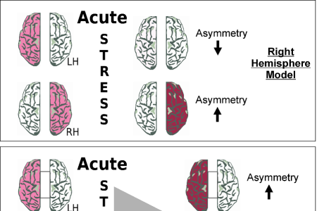

Functional hemispheric asymmetries can vary over time and steroid hormones have been shown to be one of the factors that can modulate them. Research into this matter has mainly focused on sex steroid hormones, but evidence is accumulating that stress steroid hormones might also affect asymmetries. In a newly published article, a team of researchers from the Biopsychology and Cognitive psychology labs at Ruhr-University, as well as from the University of Muenster and Utrecht University now analyzed studies in humans and animal species investigating the relation of stress and laterality. Results indicate a dual relationship of the two parameters. Both acute and chronic stress can affect different forms of lateralization in the human brain, often (but not always) resulting in greater involvement of the right hemisphere. Moreover, lateralization as a form of functional brain architecture can also represent a protective factor against adverse effects of stress. We hope that this article will stimulate further research on stress and laterality.
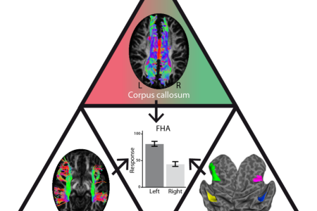

Some might think of the two brain halves as roommates - while they share the same spot within your body and like to do some things together, they still have their own interests and excel in different things. Most prominently, the left hemisphere is a genius in language whereas the right hemisphere is great in mental rotation. Researchers have often tried to find out, what causes these so called functional hemispheric asymmetries. Some argue that the answer lies in the connection between the two brain halves - others say that the difference in gray matter is key. Although both perspectives are reasonable, one factor has often been neglected: the hemispheric differences in white matter connections. In our recent review, we argue that the neurobiological underpinnings of functional hemispheric differences can only be understood, if all known factors will be combined. We hence propose a “triadic” model of functional hemispheric asymmetries, which pillars represent the differences in gray matter, the callosal connection between the two hemispheres and finally the differences in the white matter within each hemisphere. These anatomical predictors are independent from one another and deliver unique contribution in predicting functional hemispheric asymmetries. Thus, we hope to increase our understanding of this interesting phenomenon, by integrating our knowledge of different factors, instead of looking at each one separately.


How do we understand the emotional undertone (prosody) of a conversation? It is known that humans typically combine linguistic and nonlinguistic information to comprehend emotions. But how important is the contribution of prosody in the identification of emotions? To find an answer to this question, scientists from Belgium, Germany, and Austria joined forces and investigated how different communication channels interact in the identification of emotions. In the first experiment they presented their subjects synonyms of “happy” and “sad” that were spoken with either happy and sad voice. Participants had more difficulty ignoring prosody than ignoring verbal content. Thus, prosody ran strong. In the second experiment, synonyms of “happy” and “sad” were spoken with happy and sad prosody, while happy or sad faces were displayed. As expected, accuracy was low in the incongruent condition. The power of prosody became evident when participants were required to focus on verbal content only with the facial information congruent with the verbal content. Even under this condition, a discrepancy with prosodic information strongly biased the identification of emotion. Thus, the impact of prosody is unexpectedly strong in the communication of emotion. Feelings during conversation are much more than words and faces.
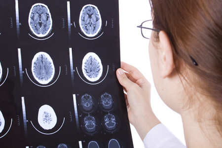

Imagine that you undergo training as a neurologist and have to group MR-images into those with and those without a tumor. Once you spot a tumor, you could simple memorize this image, and could decide on future images based on the similarity to the first tumor you saw. This way to categorize tumors is called “exemplar”-based categorization. But you could also derive an abstraction from your first tumor (its general properties) and use this summary representation for your future diagnosis. This strategy would be a “prototype”-based categorization. Subjects tested in such tasks often show different learning curves for prototype and exception stimuli. They first learn based on prototypes and then learn odd stimuli based on exemplar strategies. Neuro- and biopsychologists from Bochum now examined the contributions of different brain structures to prototype and exemplar-based category learning using functional magnetic resonance imaging (fMRI). Indeed, their subjects showed an initially superior performance for prototypes and then switched to an exemplar-based categorization for exceptions in the later learning phases. Analysis of the functional imaging data revealed that the left fusiform gyrus was involved in processing of prototypes while an activation of the right hippocampus correlated with processing of exceptions. Thus, successful prototype- and exemplar-based category learning is associated with activations of complementary neural substrates that constitute object-based processes of the ventral visual stream and their interaction with unique-cue representations, possibly based on sparse coding within the hippocampus.


Do you remember how Mowgli was dancing with Baloo on the floor of the Indian rain forest? Can you imagine one of the immortal cartoons when Asterix and Obelix where returning to their village from one of their victorious battles against the Roman army? All of these scenes have one aspect in common: The smaller person is drawn on the left, creating an ascending object sequence from left to right. After encountering much similar visualizations, psychologists from the Izmir University of Economics and from the Ruhr University Bochum pondered about the possibility that this phenomenon could be explained by the mental number line. In the mental number line numbers are represented along a left-to right-oriented continuum. Consequently, smaller object are placed on the left, creating an ascending size order from left to right. To test this idea, sixty-four participants were instructed to imagine stimulus-pairs that were staggered from those showing very prominent intra-pair size differences (e.g., elephant vs. mouse) to very low size differences (e.g., orange vs. apple). Indeed, pairs of objects were imagined with the smaller one being placed on the left. In addition, the tendency to imagine the bigger object on the right side increased directly proportional to the size difference of the two stimuli. Such a bias was also present with numbers such that the participants imagined smaller and larger numbers on the left and the right sides, respectively. Together, these findings could imply that the left-to-right orientation observed in imagined objects may share the same cognitive mechanism as the mental number line. Thus, the fact that Baloo is dancing on right side of the jungle and that Obelix walks on the right side of Asterix could reflect a common cognitive mechanism that biased the imagination of the various artists who sketched these immortal scenes with a specific orientation.
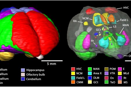

Because of their sophisticated vocal behaviour, their social nature, their high plasticity and their robustness, starlings have become an important model species that is widely used in studies of neuroethology of song production and perception. Since magnetic resonance imaging (MRI) represents an increasingly relevant tool for
comparative neuroscience, a 3D MRI-based atlas of the starling brain becomes essential. Using multiple imaging protocols, scientists from the University of Antwerp in Belgium, Université Rennes in France, and from the Biopsychology Department of the RUB delineated several sensory systems as well as the song control system. This starling brain atlas can easily be used to determine the stereotactic location of identified neural structures at any angle of the head. Additionally, the atlas is useful to find the optimal angle of sectioning for slice experiments, stereotactic injections and electrophysiological recordings. The starling brain atlas is freely available for the scientific community.

Unbelievable but true: Sarah Starosta has left the lab for a postdoc stay in New York. Sarah was an indispensable part of the Biopsychology since time being. It is very, very sad to part, but the lab of Adam Kepecs in New York is a great place to be and Sarah will certainly make the next big academic leap there. Leaving Bochum to whatever place is always sort of descend. But leaving for New York is certainly among the more acceptable options.
Hey Sarah: We will miss you a lot!!!!
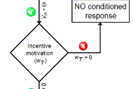

Learning and motivation are two psychological processes allowing animals to form and express Pavlovian associations between a conditioned stimulus (CS) and an unconditioned stimulus (UCS). However, most models have attempted to capture the mechanisms of learning while neglecting the role that motivation (or incentive salience) may actively play in the expression of behaviour. There is now a body of neurobehavioural evidence showing that incentive salience represents a major determinant of Pavlovian performance. This article presents a motivational model of sign-tracking behaviour whose aim is to explain a wide range of behavioural effects, including those related to partial reinforcement, physiological changes, competition between CSs, and individual differences in responding to a CS. In this model, associative learning is assumed to determine the ability to produce a Pavlovian conditioned response rather than to control the strength and the quality of that response. The model is in keeping with the incentive salience hypothesis and will therefore be discussed in the context of dopamine’s role in the brain.
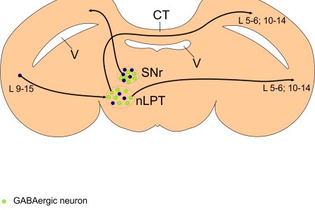

Previous studies have demonstrated that the optic tecta of the left and right brain halves reciprocally inhibit each other in birds. In mammals, the superior colliculus receives inhibitory γ-aminobutyric acid (GABA)ergic input from the basal ganglia via both the ipsilateral and the contralateral substantia nigra pars reticulata (SNr). This contralateral SNr projection is important in intertectal inhibition. Because the basal ganglia are evolutionarily conserved, the tectal projections of the SNr may show a similar pattern in birds. Therefore, the SNr could be a relay station in an indirect tecto–tectal pathway constituting the neuronal substrate for the tecto–tectal inhibition. To test this hypothesis, neuroscientists from the Biopsychology performed bilateral anterograde and retrograde tectal tracing combined with GABA immunohistochemistry in pigeons. Suprisingly, they found out that the SNr has only ipsilateral projections to the optic tectum, and these are non-GABAergic. Inhibitory GABAergic input to the contralateral optic tectum arises instead from a nearby tegmental region that receives input from the ipsilateral optic tectum. Thus, a disynaptic pathway exists that possibly constitutes the anatomical substrate for the inhibitory tecto–tectal interaction. This pathway likely plays an important role in attentional switches between the laterally placed eyes of birds.
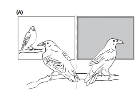

In this issue of Trends in Cognitive Sciences, Onur Güntürkün and Thomas Bugnyar discuss how the brains of birds and mammals evolved in parallel for about 300 million years to achieve comparable cognitive abilities despite quite different neural architectures, most notably the lack of an avian cortex. They present anatomical and functional evidence that the avian pallium serves the functional role of the layered mammalian cortex. Finally, they discuss how the commonalities between avian and mammalian circuits may reveal the necessary ingredients to support intelligent behavior.
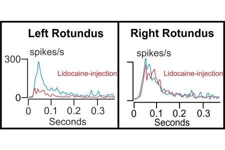

In pigeons, the left hemisphere is associated with the ability to categorize objects based on experience-based behaviors. Why is this so? To find an answer to this question, biopsychologists from Bochum and Tübingen analyzed in detail the descending pathways from the visual forebrain in pigeons. The logic behind this study is the following: If top-down control would be more efficient in the left half brain, experience-based knowledge of the forebrain could modulate left-sided ascending visual information already at thalamic level. To test this idea, single units were recorded from the visual thalamic nucleus rotundus on the left and the right side while the eyes were activated with light flashes. During holding a single unit, the visual forebrain was anesthetized with lidocaine in one hemisphere, thereby blocking top-down influences. Indeed, activity patterns of rotundal neurons were differently modulated by left and right hemispheric descending systems. Thus, visual information ascending towards the left hemisphere was modulated by forebrain top-down systems at thalamic level, while right thalamic units were strikingly less modulated. This asymmetry of top-down control could promote experience-based processes within the left hemisphere, while biasing the right side towards stimulus-bound response patterns. In a subsequent behavioral task the scientists tested the possible functional impact of this asymmetry. Under monocular conditions, pigeons learned to discriminate color pairs, so that each hemisphere was trained on one specific discrimination. Afterwards the animals were presented with stimuli that put the hemispheres in conflict. Response patterns on the conflicting stimuli revealed a clear dominance of the left hemisphere. Transient inactivation of left hemispheric top-down control reduced this dominance while inactivation of right hemispheric top-down control had no effect on response patterns. Functional asymmetries of descending systems that modify visual ascending pathways seem to play an important role in the superiority of the left hemisphere in experience-based visual tasks.
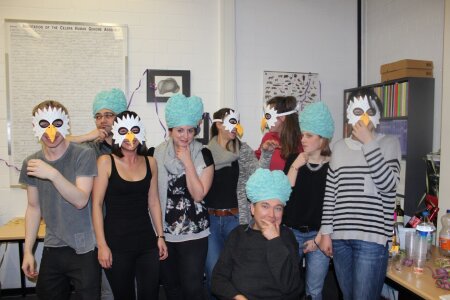

A brand-new generation of PhDs has started in the Biopsychology. Some are already with us since a short while, some will start very soon. But by and large, they form a new cohort – and they are all eager to come up with outstanding discoveries in the next couple of years. On Friday, the 19th of February 2016 they threw a great party to mark their appearance. Some of the novice PhDs will work with pigeons; others will explore the human brain. Just guess who is doing what…


Migratory birds can navigate over tens of thousands of kilometers with accuracy unobtainable for human navigators. To do so, they obviously use their brains. In this review, Biologists from Oldenburg and Biopsychologists from Bochum address how birds sense navigation- and orientation-relevant cues and where in their brains each individual cue is processed. When little is currently known, they make educated predictions as to which brain regions could be involved. They ask where and how multisensory navigational information is integrated and suggest that the hippocampus could interact with structures that represent maps and compass information to compute and constantly control navigational goals and directions. The team also suggests that the caudolateral nidopallium could be involved in weighing conflicting pieces of information against each other, making decisions, and helping the animal respond to unexpected situations. Considering the gaps in current knowledge, some of their suggestions may be wrong. However, the main aim of this theory-driven review is to stimulate further research in this fascinating field.
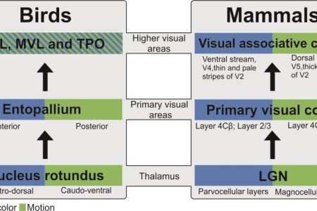

Birds show remarkable visual abilities that surpass most of our visual psychophysiological abilities. In this study, scientists from the biopsychology investigated visual associative areas of the tectofugal visual system in pigeons. Similar to the condition in mammals, ascending visual pathways in birds are subdivided into parallel form/color vs. motion streams at the thalamic and primary telencephalic level. However, we know practically nothing about the functional organization of those telencephalic areas that receive input from the primary visual telencephalic fields. The current study therefore had two objectives: first, to reveal whether these visual associative areas of the tectofugal system are activated during visual discrimination tasks; second, to test whether separated form/color vs. motion pathways can be discerned among these association fields. To this end, pigeons were trained to discriminate either form/color or motion stimuli. The immediate early gene protein ZENK was used to capture the activity of the visual associative areas during the task. Indeed, several visual associative telencephalic structures could be identified by activity pattern changes during discriminations. However, none of these areas displayed a difference between form/color vs. motion sessions. The presence of such a distinction in thalamo-telencephalic, but not in further downstream visual association areas opens the possibility that these separate streams converge very early in birds, which possibly minimizes long-range connections due to the evolutionary pressure toward miniaturized brains.

When memories are recalled, they enter a transient labile phase in which they can be impaired or enhanced followed by a new stabilization process termed reconsolidation. Meanwhile, we have more information on the neural processes that constitute this constant updating of memories stored in the synaptic weights of our brain. But we have multiple memory systems and some of them do not reside in the nervous system. It is unknown, however, whether reconsolidation is restricted to neurocognitive processes or can also be found in peripheral physiological functions as well. To answer this question, a group of psychoimmunologists from Essen and biopsychologists from Bochum used a paradigm of behaviorally conditioned taste aversion in rats to study memory-updating in learned immunosuppression. The administration of sub-therapeutic doses of the immunosuppressant cyclosporin A together with the conditioned stimulus (CS/saccharin) during retrieval blocked extinction of conditioned taste aversion and learned suppression of T cell cytokine (interleukin-2; interferon-c) production. This conditioned immunosuppression is of clinical relevance since it significantly prolonged the survival time of heterotopically transplanted heart allografts in rats. Collectively, these findings demonstrate that memories can be updated on both neural and behavioral levels as well as on the level of peripheral physiological systems such as immune functioning. A huge wide door has been opened by these insights. Memory updating seems to be a widespread process that is in no way restricted to our brain.
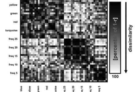

Pigeons are well capable to categorize visual stimuli. Now scientists of the biopsychology adopted a reverse engineering approach to study categorization learning in a novel way. Instead of training pigeons on predefined categories, they simply presented stimuli and analyzed neural output in search of categorical clustering on a solely neural level. They presented artificial, easily distinguishable colored shapes and grating while recording from the nidopallium frontolaterale (NFL), a higher visual area in the avian brain. They computed representational dissimilarity matrices to reveal categorical clustering based on the neural data. This revealed that colored shapes and gratings were differentially represented in the brain. This study gives proof-of-concept that this reverse engineering approach – namely reading out categorical information from neural data – can be quite helpful in understanding the neural underpinnings of categorization learning.
Most animal and human behaviors emanate from goal-directedness and pleasure seeking, suggesting that they are primarily under conscious control. However, ‘wanting’ and ‘liking’ are believed to be adaptive core subcortical processes working at an unconscious level and responsible for guiding behavior towards appropriate rewards. Here we examine whether ‘wanting’ is an inherent property of conscious goals and ‘liking’ an intrinsic component of conscious feelings. We argue that ‘wanting’ and ‘liking’ depend on mechanisms acting below the level of consciousness, explaining why individuals often struggle to enhance or refrain their motivations and emotions by means of conscious control. In particular, hyperreactivity of subcortical ‘wanting’ systems has been tied to pathological behaviors such as drug addiction and gambling disorder. In addicts, cognitive processes intended to curb drug-seeking wage a constant battle against subcortical urges to take more drug that often ends in relapse following repeated assaults. Nevertheless, we suggest that in non-pathological contexts, ‘wanting’ and ‘liking’ interact with major cognitive processes in order to guide goal-directed actions.
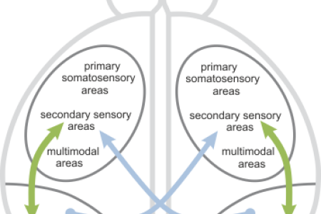

In birds which do not possess a corpus callosum the anterior commissure (AC) constitutes the main interhemispheric pathway at telencephalic level. However no detailed description of the topographic organization of the AC has been performed till now. This information is not only necessary for a better understanding of interhemispheric transfer in birds, but also for a comparative analysis of the evolution of commissural systems in the vertebrate classes. Therefore researchers from the Biopsychology Department examined the fiber connections of the AC. The main differences in the interhemispheric connectivity between birds and mammals are found at two levels of structural organization. First, the AC in birds differs from the corpus callosum and the AC of mammals in its proportion of homotopic reciprocal to heterotopic unidirectional projections. In contrast to the situation in mammals, in birds only a small amount of cells interconnect the two hemispheres in a homotopic and reciprocal fashion. Instead, most of the cells project heterotopically and in unidirectional manner. Second, in birds the absolute majority of pallial areas do not participate by themselves in interhemispheric exchange. Instead, a rather small cluster of cells is key for commissural interactions. Thus, the colloquial statement that birds are “natural split-brains” is wrong, when the pallial areas are considered that interhemispherically interact via the AC. It is true, however, when taking into account how small the proportion of pallial neurons is that constitutes interhemispheric exchange.
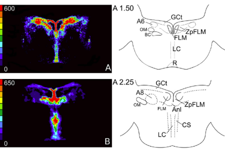

Serotonin 1A receptors play a key role in eating and sleeping. Their combined action explains why we get sleepy after a good meal. But what is the circuitry of this system? To analyze that in birds, scientists from Brazil and Germany (Bochum and Düsseldorf) joint forces and revealed the distribution of 5-HT1A receptors in the hypothalamus and brainstem of birds and analyzed their potential roles in sleep and feeding. 5-HT1A receptors are concentrated in brainstem areas like periventricular hypothalamus, preoptic nuclei and circumventricular organs that receive dense serotonergic projections. Delivering of ventricular serotonin produced a complex pattern of neuronal activations in the 5-HT1A receptor-enriched preoptic hypothalamus and the circumventricular organs, which are related to drinking and sleep regulation, but only modestly affected the activity of serotonergic neurons themselves. The same procedure induced feeding and sleeping. Further detailed experiments revealed that serotonin induces feeding and sleeping by presynaptic binding to 5-HT1A receptors that are localized in periventricular diencephalic circuits.
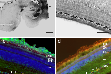

The visual system of adult pigeons shows a lateralization of object discrimination with a left hemispheric dominance on the behavioural, physiological and anatomical levels. The crucial trigger for the establishment of this asymmetry is the position of the embryo inside the egg, which exposes the right eye to light falling through the egg shell. As a result, the right-sided retina is more strongly stimulated with light during embryonic development. However, it is unknown how this embryonic light stimulation is transduced to the brain as the classic photoreceptors, rods and cones, are not yet functional. Scientists from the biopsychology department now identified a photoreceptive protein inside the retina of pigeons which is also expressed during the critical phase of asymmetry induction. This protein, Cryptochrome 1b, is expressed in retinal ganglion cells. A tracing study revealed that such Cryptochrome 1b containing ganglion cells project to the optic tectum, a primary visual area in the pigeon brain. The projection pattern and the presence of Cryptochrome 1b during the critical phase of asymmetry induction suggests that Cryptochrome 1b could indeed be the missing photoreceptive instance responsible for inducing asymmetries in the visual system of pigeons.
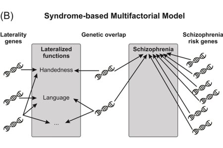

Atypical lateralization is observed significantly more often in schizophrenia patients than in the general population, which led several authors to conclude that there is a genetic link between laterality and schizophrenia. However, the molecular genetic evidence for a link between laterality and schizophrenia is weak. In the present article, a multinational team of researchers from the Biopsychology lab and the Bergen fMRI Group in Norway reviewed recent genetic evidence indicates that schizophrenia is not a single disorder but a group of heritable disorders caused by different genotypic networks leading to distinct clinical symptoms. Based on these findings, the researchers suggested a new theoretical framework in which genetic favtors are not mapped on schizophrenia as a whole but on discrete schizophrenia symptoms.


People differ in the capacity to regulate emotions, thoughts and behaviors which are directed towards fulfilling one’s intentions. We differentiate between two personality types, termed action-orientation and state-orientation. Action oriented individuals are very efficient in pursuing their intentions. They focus on the relevant information and have no difficulties in taking the necessary steps towards their goal. Yet, state oriented individuals exhibit difficulties in these so called action control strategies. They tend to perseverate on irrelevant information, hindering them to pursue their intentions. This is why these two types are colloquially referred to as “doers” and “brooders”. While it is known that these behavioral differences exist, it is not clear as to whether they also reflect a neuronal deficit in state oriented individuals. Our results revealed that action oriented individuals inhibit more easily irrelevant information than state oriented individuals and also exhibit a different neuronal activity. Compared to state oriented individuals, action oriented individuals displayed a shorter latency of the frontocentral N2, an ERP component that reflects inhibition and cognitive control. These results show for the first time that neuronal processes are accountable for the incapacity of state oriented individuals in pursuing their goals more efficiently. Furthermore, they indicate that therapeutic interventions in different fields (e.g. overweight, addiction) would profit from a more individual perspective. While some strategies might be very successful for action oriented individuals, they might not be so for state oriented individuals.
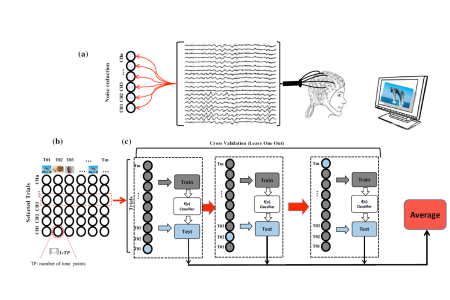

Oscillations of electroencephalographic (EEG) signals represent the neural activity which reflecting a mental state or cognitive process that arise from the behavioral task and sensory representations across the mental state activity. Previous studies have shown the relation between event-related EEG and sensory-cognitive representation, and have revealed that categorization of presented objects can be successfully recognized using recorded EEG signals when subjects view objects. In this study, we utilized the recording of EEG signals in conjunction with a multivariate pattern recognition technique with the aim of identifying neural activity associated with conceptual representation based on the presentation of semantic categories of objects. Using multivariate stimulus decoding methods, surprisingly, they demonstrated that object discrimination activity is apparent from the phase pattern of EEG signals across the time in low frequency bands (1-4 Hz), but not by the power of oscillatory brain signals in the same frequency band. In contrast, discrimination accuracy from the power of EEG signals has significantly higher than the performance by the phase of EEG signal in the high frequency band (20-30 Hz). Moreover, our results indicate that how the accuracy of prediction changes between various areas of the brain continuously across the time. In particular, we found that, during the object categorization task, the inter-trial phase coherence (IPC) in low frequency bands is significantly higher than other frequencies in various regions of interests. This measure is associated with decoding pattern across the time. These results suggest that the mechanism underlying conceptual representation can be mediated by the phase of oscillatory neural activity.
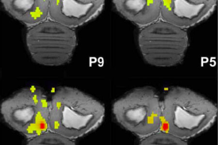

Functional hemispheric asymmetry is a common feature of brain organization, yet little is known about how hemispheric dominance is implemented at the neural level. Birds have a strong left-hemisphere asymmetry in navigation. Since the hippocampus plays a key role in spatial orientation, a team of biopsychologists from Bochum as well as Belgian and US American neuroscientists set out to test, if left and right hippocampi have asymmetrically organized hippocampal networks in birds. For this to reveal, they relied on resting state fMRI analyses in awake and sedated birds in scanners with extremely high magnetic field strengths. Indeed, the could show that following seeding in either an anterior or posterior region of the hippocampal formation of homing pigeons and starlings, the emergent functional connectivity maps are consistently larger following seeding of the left hippocampus. Left seedings were also more likely to result in networks that extend to the contralateral hippocampus and outside the boundaries of the hippocampus. The data support the hypothesis that broader functional connectivity is one neural-organizational property that confers, with respect to navigation, a functional dominance to the avian left hippocampus.
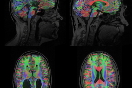

Previous research indicates that callosal supression is important for the establishment of functional brain asymmetries. In this study researchers from the Biopsychology lab wondered what whould happen to callosal supression when there is no corpus callosum? They tested this in a multimethod neuroimaging approach combining functional and structural connectivity analysis in patients who were born without a corpus callosum and healthy individuals. The study revealed two important features of the corpus callosum. First, in healthy individuals, interhemispheric motor inhibition measured with fMRI is related to properties of specific motor fibers in the corpus callosum. Second, the inhibitory interaction between motor areas is diminished in patients without a corpus callosum. Interestingy, for these patients motor areas in each hemisphere showed symmetric involment in motor functions, indicating a reduced functional brain asymmetry. These results provide novel insights into callosal functions and the establishment and maintenance of functional brain asymmetries in general.


Current Biology has a nice tradition: From time to time they feature a scientist on a full page by asking a lot questions (some of them strange). In the issue of October 19, 2015, it was Onur Güntürkün’s turn: “Do you have heroes?” (Yes, many); “If you would not have made it as a scientist, what would you have become?” (A taxi driver); “What is the best advice you’ve been given?” (You better give up the science you do), etc.
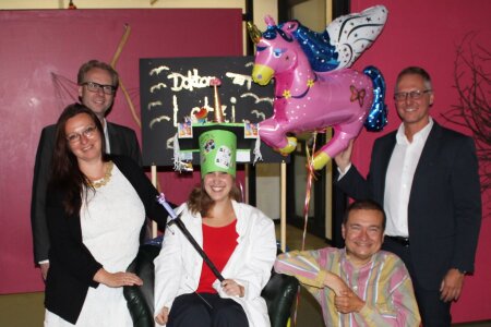

On Thursday, the 3rd of September 2015, Sarah defended her PhD thesis entitled „„Einflüsse ontogenetischer Faktoren und kommissuraler Systeme auf die asymmetrische Informationsverarbeitung in der Taube (Columba livia)“. After giving a brilliant presentation, she stood the tough questions of the examination committee. Her thesis was a great mixture of neuroanatomical and behavioral studies. After close to an hour of questions, the committee unanimously decided that she had done a great job and decided to award her the title of a Dr. rer. nat. After that, Sara was dragged as “Princess of Biopsychology” on her own feudal cart through the whole building. Afterwards, Her Highness invited everybody to party.
Congratulations Sara! We are proud of you!

Whistled languages represent an experiment of nature to test the widely accepted view that language comprehension is governed by the left hemisphere in an input-invariant manner. Indeed, the left hemisphere does the job for writing as well as atonal, tonal, signed, and clicked languages. The right hemisphere, on the other hand, is specialized to encode acoustic properties like spectral cues, pitch, and melodic lines and plays a role for prosodic communication. Would left hemispheric superiority change, when subjects had to encode a language that is constituted by acoustic properties for which the right hemisphere is specialized? Well, whistled Turkish uses the full lexical and syntactic information of vocal Turkish, and transforms this into whistles to transport complex conversations over kilometers. A group of Biopsychologists from Bochum now tested the comprehension of vocally vs. whistled syllables in native whistle-speaking people of mountainous Northeast Turkey. They discovered that whistled language comprehension relies on symmetric hemispheric contributions, associated with a decrease of left and a relative increase of right hemispheric encoding mechanisms. These results demonstrate that a language that places high demands on right hemisphere-typical acoustical encoding mechanisms creates a radical change in language asymmetries. Thus, language asymmetry patterns are importantly shaped by the physical properties of the lexical input. This paper was featured by many prime media like Science, Scientific American, The Scientist, New York Times, The New Yorker, BBC, Washington Post, CNN, and many more. It is also part of the Science podcast of August 21, 2015.
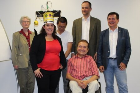

On Thursday, the 13th of August 2015, Sarah defended her PhD thesis entitled „Neuronal and Behavioral Mechanisms of Extinction Learning and Renewal” in front of a tough IGSN examination committee. One of the examiners, Juan Rosas, had travelled all the way from Spain to Bochum for this task. As expected, Sarah did a brilliant job. Her talk was simply perfect, and she could respond in excellent and witty ways to each and every question. Despite trying in the hardest possible way, the examiners found no soft spot. It was all simply perfect. Therefore, the committee unanimously decided to award her the title Philosophiae doctoris (PhD) in Neuroscience.
Congratulations Sarah! You are outstanding!


Die Habilitation ist die höchstrangige Hochschulprüfung in Deutschland und setzt ein exzellentes wissenschaftliches Oeuvre voraus. Nach einem eingehenden Prüfverfahren mit auswärtigen internationalen Gutachten wird bei erfolgreicher Evaluation die Lehrbefähigung (facultas docendi) vergeben. Nach einer anschließenden erfolgreich verlaufenden Antrittsvorlesung wird zum Schluss die Lehrberechtigung ausgesprochen. Diese heißt venia legendi („Erlaubnis zu lesen [d. h. zu lehren]“). Die venia legendi ist die höchste Auszeichnung, die eine Fakultät vergeben kann und signalisiert den Abschluss eines Prüfprozesses mit dem evaluiert wird, ob der Wissenschaftler sein Fach in voller Breite in Forschung und Lehre vertreten kann. Bei Erfolg wird die akademische Bezeichnung Privatdozent (PD) verliehen. Am 8. Juni 2015 war es für Sebastian Ocklenburg soweit. Mit seinem Vortrag „Das asymmetrische Gehirn und die Doppelhelix - Die Neurogenetik funktionaler Lateralisation" überzeugte er restlos alle, dass er diesen Titel verdient hatte und bekam die venia legendi aus den Händen von Annette Kluge, der Dekanin in spe unserer Fakultät.
Herzlichen Glückwunsch PD Dr. Sebastian Ocklenburg!!!!
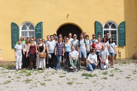

Towards the end of July the Biopsychology lab departed for its first grand retreat towards Bavaria. On the first morning we enjoyed the impressive research station of our host Princess Auguste von Bayern in her castle Leutstetten. Her outdoor aviaries and her insights in Social learning and Tool use of her New Caledonian Crows fascinated us as much as her heartfelt kindness. The picture was taken in front of her castle. Then came the excursion to the Max-Planck-Institute for Ornithology with lectures of Manfred Gahr, Nils Rattenborg, Henrik Brumm and Holger Görlitz. We could use their excellent facilities on Saturday for our own series of presentations, showing up future ideas and synergies in our department. The last day was spent as a guest of Auguste's father on the Ritterfestival where some of us unfortunately were sentenced to tarring and feathering. A somehow fitting punishment for members of a lab working with birds...
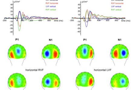

While it is well known that the left hemisphere is more efficient than the right in most tasks involving perception of speech stimuli, the neurophysiological pathways leading to these lateralised performance differences are as yet rather unclear.
To clarify this question, a team of researchers from the University of Dresden and the Biopsychology lab recorded event-related potentials (ERPs) during tachistoscopic presentation of horizontally or vertically presented verbal stimuli in the left (LVF) and the right visual field (RVF). On the behavioural level, participants showed stronger hemispheric asymmetries for horizontal, than for vertical stimulus presentation. In addition, ERP asymmetries were also modulated by stimulus presentation format.
Moreover, sLORETA revealed that ERP left-right asymmetries were mainly driven by the extrastriate cortex and reading-associated areas in the parietal cortex. Taken together, the present study shows electrophysiological support for the assumption that language lateralisation during speech perception arises from a left dominance for the processing of early perceptual stimulus aspects.

Anlässlich des „Deutsch-Türkischen Wissenschaftsjahres 2014-15" haben die Leopoldina und die Humboldt-Stiftung ein gemeinsames Format ins Leben gerufen – die „Deutsch-Türkischen Wissenschaftsgespräche" ("German-Turkish Science Dialogue"). Ziel ist es, den bilateralen Dialog zu fördern und die deutsch-türkische Wissenschaftskooperation zu unterstützen. Die Leopoldina nimmt als Nationale Akademie der Wissenschaften Deutschlands mit ihren rund 1500 Mitgliedern zu den wissenschaftlichen Grundlagen politischer und gesellschaftlicher Fragen unabhängig und öffentlich Stellung. Sie vertritt die deutsche Wissenschaft in internationalen Gremien und handelt zum Wohle der Menschen und der Gestaltung ihrer Zukunft. Die Alexander von Humboldt-Stiftung fördert Wissenschaftskooperationen zwischen exzellenten ausländischen und deutschen Forscher/innen.
Dienstag, 16. Juni 2015, 14:00 bis 19:00 Uhr
Ruhr-Universität Bochum, Gebäude GAFO 03 / 252, Universitätsstraße 150, 44801 Bochum
Neuroscience: From Structure to Function and Back
Eine Online-Registrierung unter www.leopoldina.org/de/science-dialogue wird bis zum 14. Juni 2015 erbeten.


Am 20. Mai wurde Prof. Dr. Dres. h.c. Onur Güntürkün als eines von 19 neuen Mitgliedern in der Nordrhein-Westfälischen Akademie der Wissenschaften und Künste in Düsseldorf willkommen geheißen. Mit seiner Aufnahme ist Prof. Güntürkün der erste Psychologe in der Klasse für Naturwissenschaften. Die AWK ist eine Vereinigung der führenden Forscher des Landes und die Heimat von 14 wissenschaftlichen Forschungsvorhaben. Sie wurde 1970 als Nachfolgeeinrichtung der Arbeitsgemeinschaft für Forschung des Landes Nordrhein-Westfalen gegründet.
© AWK NRW/Andreas Endermann
v. l.: RUB-Rektor Prof. Dr. Elmar Weiler, Prof. Dr. Käte Meyer-Drawe, NRW-Wissenschaftsministerin Svenja Schulze, Prof. Dr. Onur Güntürkün und Prof. Dr. Dr. Dr. Hans Hatt.


Martina Manns becomes an Editorial Board Member for Scientific Reports. This journal is a young online, open access journal from Nature Publishing Group. It publishes scientifically valid primary research from all areas of the natural and clinical sciences and is already the 5th among all multidisciplinary research journals. Martina will bring in her expertise in Cognitive Neuroscience and Developmental Biopsychology to ensure publication of scientifically valid and technically sound research.


Wir sind unser Gehirn; es steuert unsere Handlungen und Gefühle. In 4 DVDs und einem Begleitbuch mit Abbildungen und weiterführender Literatur gibt Onur Güntürkün einen weit gefächerten Einblick in die Hirnforschung. Mit vielen Experimenten führt er die Zuschauer in 10 Kapiteln durch die Neurowissenschaft. Am Ende jeder Lektion diskutieren dann ZEIT-Redakteur Ulrich Schnabel und Onur Güntürkün über die Implikationen dieser Befunde. Zuerst wird der Aufbau des menschlichen Gehirns erläutert und das Prinzip der neuralen Übermittlung erklärt. Dann werden Fragen besprochen wie z. B.: Wie nehmen wir die Welt wahr? Wie lernen und wieso vergessen wir? Was ist Intelligenz und lassen sich Gedanken steuern? Wie und wieso erleben wir Emotionen? Unterscheidet sich das Denken von Männern und Frauen und wenn ja, warum? Warum haben unsere Gehirnhemisphären unterschiedliche Funktionen? Diese Inhalte werden mit Erkenntnissen der Störungen des Gehirns, sowie seiner Veränderung während der gesamten Lebensspanne verknüpft.
http://www.zeitakademie.de/seminare/koerper-geist/hirnforschung?gclid=CL2bhf6b0sUCFezKtAodMwoAIQ


Birds can rely on a variety of cues for orientation during migration and homing. Celestial rotation provides the key information for the development of a functioning star and/or sun compass. This celestial compass seems to be the primary reference for calibrating the other orientation systems including the magnetic compass. Thus, detection of the celestial rotational axis is crucial for bird orientation. Now, Biologists from Oldenburg and Biopsychologists from Bochum used operant conditioning to demonstrate that homing pigeons can principally learn to detect a rotational centre in a rotating dot pattern and examined the behavioural response strategies of pigeons in a series of experiments. Initially, most pigeons applied a strategy based on local stimulus information such as movement characteristics of single dots. One pigeon seemed to immediately ignore eccentric stationary dots. After special training, all pigeons could shift their attention to more global cues, which implies that pigeons can learn the concept of a rotational axis. The ability to precisely locate the rotational centre was strongly dependent on the rotational velocity of the dot pattern and it crashed at velocities that were still much faster than natural celestial rotation. The study thus suggest that the axis of the very slow, natural, celestial rotation could be perceived by birds through the movement itself, but that a time-delayed pattern comparison should also be considered as a very likely alternative strategy.


Imagine a regular sequence of sounds: ba – ba – ba – ba- ba… And then think of an odd sound in between: ba – ba – ta – ba- ba… Neuroscientists will record a strange evoked potential from your auditory cortex as soon as you hear the “ta”. This potential is known as mismatch negativity (MMN) and it depends on the activation of NMDA-receptors. MMNs were recorded from the brains of humans, other primates and rodents and are seen as a cortex-specific neural component that guides attention switches to changes in the environment. Now, psychiatrists from Newcastle (Australia) and Biopsychologists from Bochum investigated if an MMN-like potential can also be recorded from the auditory-cortex equivalent forebrain structure in pigeons. Indeed, an MMN-like field potential was recorded and blocked with NMDA-receptor blocker. These results are suggestive of similar auditory sensory memory mechanisms in birds and mammals that are either homologue from a common ancestor 300 million years ago or resulted from convergent evolution.


Last week Patrick Anselme departed from Belgium and arrived with his own DFG-grant in Bochum to start his research project. So, what is he planning to do? His research agenda starts with a simple observation: In order to survive, humans and other animals must maximise energy intake. However, sometimes strange things happen: animals start preferring an unpredictable food option which provides a lower reward rate. Thus, they gamble; But why? Do animals sometimes prefer the unpredictable since they might obtain reward sooner? Or do they try to maximize reward? In fact, both factors might be combined in gambling: individuals try to maximize gains, and unpredictability keeps their interest upright. In his project, Patrick plans to understand if the motivation to maximize reward and the unpredictability of reward can control choices in pigeons. In addition, he plans to see how dopamine determines sensitivity to these two factors since dopamine is chiefly recruited by rewards, but is also sensitive to unpredictability.
Good luck, Patrick, and welcome to the Biopsychology!!
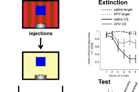

Within the Forschergruppe 1581 “Extinction learning” we conducted a further experiment on context-dependent extinction learning under appetitive conditions. The results are now published within the special Frontiers Research Topic “Extinction learning form a mechanistic and systems perspective”. We investigated the responsibility of N-methyl-D-aspartate receptors (NMDARs) within the `prefrontal` caudal nidopallium (avian functional equivalent of mammalian prefrontal cortex) for contextual extinction learning.
The ability to flexibly adapt to new contingencies by learning and to inhibit previously acquired associations in a context-dependent manner is essential for extinction learning. To uncover neuronal network beyond the bench of aversive extinction learning we reused the previous establish sign tracking within-subject ABA-renewal paradigm (Lengersdorf et al, 2014). Here we shed light on invariant properties of the neural basis of extinction learning by employing the pigeon as a model system. Since NMDARs in prefrontal cortex have been shown to be relevant for extinction learning, we locally antagonized NMDARs through 2-Amino-5-phosphonovalerianacid (APV) during extinction learning. This slowed down extinction learning and in addition caused a disinhibition of responding to a continuously reinforced control stimulus. In subsequent retrieval sessions, spontaneous recovery was increased while ABA renewal was unaffected. The effect of APV resembles that observed in studies of fear extinction with rodents, suggesting common neural substrates of extinction under both appetitive and aversive conditions and highlighting the similarity of mammalian prefrontal cortex and the avian caudal nidopallium despite 300 million years of independent evolution.

Left-right asymmetries of structure and function are a common organization principle in vertebrates brain organization, but many aspects of this fascinating phenomenom are not well understood. Over the course of the last few years, researchers from the Biopsychology lab, the Action Lab of the University of Dresden and the Bergen fMRI group edited a large, multinatioral Research Topic in Frontiers in Psychology, encompassing peer-reviewed 34 articles from leading authorities in the field of lateralization research. With cutting-edge research in different species ranging from insects to humans, the ebook highlights many of todays main sections of lateralization research and presents a valuable ressource for researchers interested in the field.
The ebook can be downloaded here.
The question, what constitutes the neural correlates of handedness has never been answeres unequivocally. In a recent paper by Guadalupe et al. (2014) (link to the original article) the authors investigated an impressively large sample of 1960 right-handed and 106 left-handed participants to answer this question. The authors compared left- and right-handers regarding the cortical surface area of 10 different candidate regions related to language, motor control and visual processing which were obtained from previous studies investigating the structural correlates of handedness in the brain. While the authors found a nominally significant association between handedness and the surface area of the left precentral sulcus, not a single effect survived statistical correction for multiple testing. Frontiers in Psychology invited a team of lateralization experts from the Biopsychology lab to comment on this important discovery in a prestigious Frontiers Commentary Article.
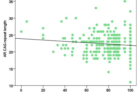

Sex differences in handedness have long been known, but their molecular basis is not well understood. In the present study, a team of scientists from the IKN and the Human Genetics Lab investigated the relationship of CAG-length variation in the androgen receptor gene AR and handedness in a large sample of over 1000 participants. Mixed-handedness in men was significantly associated with longer CAG-repeat blocks and women homozygous for longer CAG-repeats showed a tendency for stronger left-handedness. These results suggest that handedness in both sexes is associated with AR CAG-repeat length, with longer repeats being related to a higher incidence of non-right-handedness. Since longer CAG-repeat blocks have been linked to less efficient AR function, these results implicate that differences in AR signaling in the developing brain might be one of the factors that determine individual differences in brain lateralization.

Metabotropic glutamate (mGlu) receptors and, in particular, mGlu5 are crucially involved in forms of hippocampus-dependent synaptic plasticity that are believed to also underlie extinction learning. MGlu5 is also required for information transfer through neuronal oscillations and for spatial memory. This places this receptor in a unique position with regard to information encoding. Therefore, neurophysiologists and biopsychologists from Bochum explored the role of this receptor in context-dependent extinction learning under constant, or changed, contextual conditions. Their results support that although extinction learning in a new context is unaffected by mGlu5 antagonism, extinction of the consolidated context is impaired. This suggests that mGlu5 is intrinsically involved in enabling learning that once-relevant information is no longer valid.


Im Alltag ist der Mensch ständig mit Situationen konfrontiert, in denen früher Gelerntes nicht mehr gültig ist – Psychologen sprechen vom „Extinktionslernen“. Die damit verbundenen Mechanismen auf Verhaltens-, Hirn- und Immunebene am Beispiel von Tauben, Ratten und Menschen zu verstehen, ist Ziel der Forschergruppe 1581. Die Erkenntnisse dieser Initiative können langfristig auch der Therapie von Angstpatienten und Menschen mit Organtransplantationen zugutekommen. In der aktuellen Ausgabe des DFG-Magazins forschung berichtet Onur Güntürkün von den Untersuchungen innerhalb der Forschergruppe.
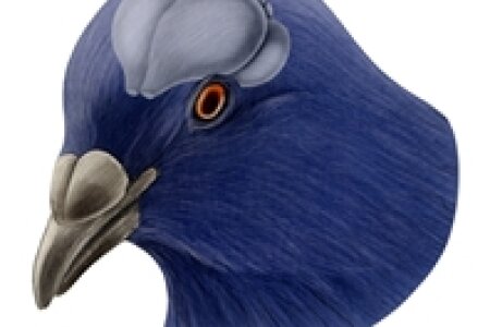

They fly over hundreds of kilometers straight to their home. They memorize hundreds of abstract symbols, work for hours on problems without any sign of fatigue, manage logical problems like transitive inference, are in several respects cognitively on par with monkeys, have an unbelievably high frustration threshold during behavioral studies… and do all this for just a handful of grain. Pigeons have remarkable asymmetries of brain function, a reasonably well-charted nervous system and breast muscles that taste so well when served with wild mushrooms and a rich red wine gravy. In a recently published paper, Biopsychologists from Bochum have concocted a review of all the merits of pigeons as experimental animals for cognitive neuroscience. A sort of memorial that was long overdue.

On Friday the 12th of December 2014 Daniel Lengersdorf defended his PhD thesis entitled „ Function of the pigeon nidopallium caudolaterale and hippocampus in context-dependent extinction learning under appetitive conditions”. At its core, Daniel’s thesis opens a new path on the analysis of the possibly invariant properties of the neural fundaments of extinction learning in birds and mammals. With a set of ingeniously designed behavioral experiments, Daniel managed to show the specific contribution of the avian “prefrontal cortex” and the bird hippocampus on extinction learning and on context-dependent extinction memory retrieval. In addition, Daniel was able to show the contribution of ‘prefrontal’ NMDA-receptors for these functions. In his defense, Daniel successfully managed to answer all questions that were put forward by the committee. All in all, a great performance! Therefore, the committee unanimously decided to award him the title Dr. rer. nat. Congratulations Daniel! We are truly proud of you.

Die Neurowissenschaft ist zur Leitwissenschaft geworden. Neurowissenschaftlern wird daher vieles zugetraut. Manchmal zu vieles. Sie sollen möglichst schnell die Mechanismen des Gehirns verstehen und dadurch alle Krankheiten des Gehirns heilen; sie sollen die Organisation von Schulen „hirngerecht“ organisieren, bei der Lösung gesellschaftlicher Probleme beratend zur Seite stehen und bei vielen wissenschaftlichen Kontroversen das letzte Wort haben. Diese übertriebenen Erwartungen entstanden z. T. durch überhöhte Ankündigungen von Regierungen (z. B. „Decade of the Brain“) sowie Verlautbarungen einzelner Neurowissenschaftler in Form scheinbar wissenschaftlich gesicherter Aussagen (z. B. die Aussagen zur Willensfreiheit). Um längerfristig einem Vertrauensverlust zu begegnen, organisiert die Hertie-Stiftung zusammen mit der Frankfurter Allgemeinen Zeitung eine Vortragsreihe, bei der einmal im Monat ein Neurowissenschaftler zu einem aktuellen Thema der Hirnforschung einen öffentlichen Vortrag hält. Darin sollen die gesellschaftlichen Zuschreibungen, die tatsächliche Erkenntnislage und die möglichen wissenschaftlichen Grenzen der Neurowissenschaft bezüglich eines konkreten Themas vorgestellt werden. Am 22. Oktober 2014 hielt Onur Güntürkün an der Frankfurter Universität den Vortrag zum Thema „Denken“. Sein Vortrag mit dem Titel „Die Gedanken sind frei – aber werden sie das auch bleiben?“ erschien am 29.10.2014 in der FAZ.
Die Gedanken sind frei – aber werden sie das auch bleiben? FAZ, 29.10.2014

Sebastian Ocklenburg successfully habilitated on the 12th of November 2014 in the Faculty of Psychology for the field of “Cognitive Neuroscience”. His written thesis already had impressed everybody by its clear focus on cerebral asymmetries and its breadth within this area of analysis. Now, he performed equally well with his oral presentation on why white matter matters. Sebastian’s presentation was absolutely lucid and perfect. He could subsequently answer all emerging questions in the best possible way. Altogether, this was a very strong presentation and the committee unanimously decided that Sebastian Ocklenburg should be awarded with the habilitation of the Faculty of Psychology.
CONGRATULATIONS SEBASTIAN !!!
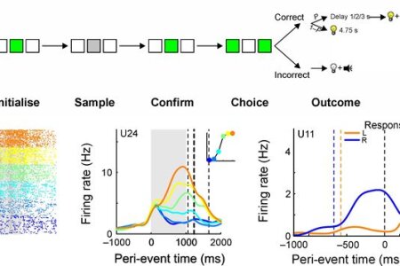

In the recently published paper we focused on the investigation of generality in neural mechanisms underlying perceptual decision making across species. Therefore, we recorded single-neuron activity in the pigeon nidopallium caudolaterale (NCL). This area is a non-laminated associative forebrain structure thought to be functionally equivalent to mammalian prefrontal cortex. The freely moving subject performed a well established visual categorisation task. Whereas the majority of NCL neurons unspecifically upregulated or downregulated activity during stimulus presentation, ~20% of neurons exhibited differential activity for the sample stimuli and predicted upcoming choices. Moreover, neural activity in these neurons was ramping up during stimulus presentation and remained elevated until a choice was initiated. This response pattern is similar to that found in monkey prefrontal and parietal cortices in saccadic choice tasks. In addition, many NCL neurons coded for movement direction during choice execution and differentiated between choice outcomes (reward and punishment). By means of these results we further implicate the NCL to be involved in the selection and execution of operant responses, an interpretation resonating well with the results of previous lesion studies. The resemblance of the response patterns of NCL neurons to those observed in mammalian cortex suggests that, despite differing neural architectures, mechanisms for perceptual decision making are similar across classes of vertebrates.


Die Frage „Wie wurde ich zu der Person, die ich bin?“ betrifft jeden ganz unmittelbar. Wenn man möglichst umfassend verstehen will, welche Bedingungen, Prozesse und Einflussfaktoren dazu beitragen, dass wir in der Interaktion mit unserer Umwelt zu einzigartigen Individuen werden, muss man das Panorama des gegenwärtigen Wissens über die natürlichen und kulturellen Wurzeln menschlicher Individualität aus den unterschiedlichsten wissenschaftlichen Perspektiven betrachten. Alte Erklärungsmodelle, die Eigenschaften und Leistungsvermögen eines Menschen entweder auf das Erbgut oder auf Umwelteinflüsse zurückführten, werden abgelöst durch neue Erkenntnisse über die Wechselwirkung dieser Faktoren. Doch wie genau resultiert der einzelne Mensch aus der Ko-Konstruktion von Geist, Gehirn, Genom und Gesellschaft? Die Leopoldina hatte ihre Jahresversammlung 2013 dieser Frage gewidmet. Unter dem Motto „Geist – Gehirn – Genom – Gesellschaft. Wie wurde ich zu der Person, die ich bin?“ haben vom 20. bis 22. September 2013 in Halle Vertreter der unterschiedlichsten Disziplinen aus Sicht ihrer Fachgebiete dargestellt, wie wir Individuen aus dem Zusammenspiel von Geist, Gehirn, Genom und Gesellschaft resultiert. Nun ist der Tagungsband erschienen, der fast alle auf der Jahresversammlung gehaltenen Vorträge enthält (Güntürkün, O., Hacker, J. (Hrsg.), Nova Acta Leopoldina Nr. 405, Band: 120, Geist – Gehirn – Genom – Gesellschaft: Wie wurde ich zu der Person, die ich bin? Wissenschaftliche Verlagsgesellschaft Stuttgart, 2014.). Der untere Link öffnet den Eröffnungsvortrag zur Jahrestagung von Onur Güntürkün.


www.dasGehirn.info provides information on the brain, its functions and its importance to our feelings, thoughts and actions - comprehensive, readable, attractive and clearly in words, pictures and sound. This time, the online magazine informs about the 49th Congress of the German Society for Psychology (DGPs) in Bochum. This year's motto was "Diversity of Psychology". At the same time the Institute of Psychology was celebrating its 50th anniversary. Learn more about envy, nuts and more and see the video here.

The online magazine www.dasGehirn.info informs the reader about findings of brain research in an entertaining and comprehensive way. In cooperation with the research groups FOR 1581 and SFB 636 they present extinction learning as one of the most exciting current research topics between mind and brain. As old knowledge is persistent - especially under stress -, the initial process of acquisition of new knowledge is well studied, but the process of extinction is far less understood. Read more.


Onur Güntürkün will stay from October 2014 to April 2015 in Berlin Grunewald together with about 50 other researchers from all over the world to think deeply about scientific problems. During this time, he wants to explore if it is possible that the capacity for complex cognition arose several times during evolution such that groups of non-mammalian animals might have developed as-yet unknown brain mechanisms that generate intelligent behavior. His ideas may contribute to developing a new theory of the evolution of brains and complex cognitive abilities.
In the meantime during WS 2014/15, Martina Manns acts for a substitute professor of the Biopsychology. Apart from taking over teaching, she will pursue investigating development of cerebral asymmetries in pigeons to unravel the interactions of left- and right-hemispheric specialization processes.
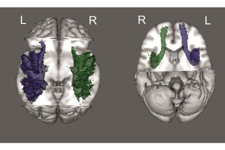

Structural asymmetries in white matter tracts within the language system have been suggested to be one of the factors underlying functional language lateralization. To test this assumption, researchers from Bergen, Norway, and the biopsychology lab in Bochum examined how performance in the dichotic listening task is affected the structure of the arcuate and uncinate fasciculus, assessed with DTI. Both arcuate and uncinate fasciculus had a larger tract volume in the left compared to the right hemisphere, but fractional anisotropy was higher in the right than in the left arcuate fasciculus. Interestingly, structural asymmetries were linked to functional lateralization, that is, tract volume and fractional anisotropy of the left arcuate fasciculus were positively correlated to the strength of functional language lateralization, as was the volume of the right uncinate fasciculus. These results suggest that both micro- and macro-structural properties of language-relevant intrahemispheric white matter tracts modulate the behavioral correlates of language lateralization.

On Friday the 11th of July 2014 Nils Kasties defended his PhD thesis entitled „Neuronal foundations of decision making in the pigeon nidopallium caudolaterale”. …And what a defense it was!!! Nils presented two major experiments and perfectly embedded them into the current literature. In the end, he was able to integrate his findings into a model of decision making in the avian forebrain. Afterwards, Nils bravely and successfully defended his arguments for a full hour against a whole barrage of counterarguments until the members of committee were exhausted and were just yearning for sparkling wine and party. But before the party could start, members of the committee unanimously decided to award Nils the title Dr. rer. nat. Congratulations Nils! We are proud of you. In the picture you can see Nils with his two advisors on his PhD-cart with which he was drawn back to the lab.

Learning is a fundamental ability of all organisms and the underlying neuronal mechanisms are highly conserved across species. To understand the mechanisms of learning at the single neuron level, we seek to record the activity of individual neurons across different learning stages. With this goal, RUB biopsychologists designed a behavioral task which encompassed three learning stages in a single experimental session. They made use of the keenness of pigeons in operant tasks who are willing to perform a vast amount of trials (>1000) for a relatively low amount of reinforcers. In addition, several methodological measurements were implemented to improve and control recording quality. To sum up and conserve the used methodological standards, we published a video article in the Journal of Visualized Experiments (JoVE), where the sophisticated behavioral paradigm as well as our methodological inventions are visualized and described in detail.


Recently, the government of India decided by law that "cetaceans...should be seen as 'non-human persons' and as such should have their own specific rights." This decision results from the political pressure, the public opinion, and the work of a small group of scientists who argue that dolphins have an intelligence that comes close to humans. In Germany activists and members of the Green Party demand that the dolphinaries in Duisburg and Nürnberg be closed. But how strong is the scientific evidence for the cognitive exceptionality of dolphins? Paul Manger [Neuroscience, 2013] reviewed the dolphin cognition literature and drew a quite sobering conclusion. But is his critique justified or does he throw the baby out with the bathwater? In an invited review to Brain, Behavior and Evolution (2014), Onur Güntürkün summarizes the literature on dolphin cognition and compares it with evidences from other animals. He concludes that dolphin cognition is not exceptional since there is not a single achievement that has not also been shown in several other species. However, in all major areas of comparative cognitive science, dolphins have been shown to achieve fast learning, high flexibility, and a swift transfer of learned knowledge to new contingencies. So, dolphins are in many respects cognitive generalists, performing at an overall high level. So, the evolution of high cognitive skills has independently taken place in several lines of life, among them primates, cetacean (mostly dolphins), birds (mostly corvids and parrots). There seem to be many different routes to intelligence.
Güntürkün, O. (2014). Is dolphin cognition special? Brain Behav. Evol., 83: 177-180.
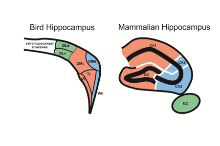

The evolutionary lines of birds and mammals diverged nearly 300 million years ago. As a consequence, the brains of these two classes of vertebrates share some features but also have many unique and non-shared properties. The hippocampus is an especially interesting case: the hippocampus of mammals and birds is homologous and thus stems from a common reptilian ancestor that already had a hippocampus. As a result, avian and mammalian hippocampi share some similar functions (e.g. spatial cognition) and anatomical features (e.g. a re-entrant loop). But they also developed a large number of bird- or mammal-typical aspects that do not seem to have counterpart. Overall, it is still mostly unknown which components of the bird hippocampus are avian-unique and which are similar to mammals. To solve these questions, scientists from Bochum, Düsseldorf, and Bowling Green (USA) analyzed binding site densities for 11 different receptor systems by using quantitative in vitro receptor autoradiography. Additionally, they labeled zinc to show a further compartmentalization of the hippocampus. The regionally different receptor densities mapped well onto seven hippocampal subdivisions previously described. Several differences in receptor expression highlighted distinct hippocampal subfields. In addition, several similarities in receptor binding densities between subdivisions of the avian and mammalian hippocampus were observed. Despite the similarities, the consortium of scientists concluded that 300 hundred million years of independent evolution has led to a vast mosaic of similarities and differences in the organization of the avian and mammalian hippocampal formation. Thinking about the avian hippocampus in terms of the strict organization of the mammalian hippocampus is likely insufficient to understand the hippocampus of birds.


Since a while, rumor had it. Then it became official; and now it truly happened: Maik Stüttgen left the Biopsychology to become a Professor for Behavioral Neurophysiology at the University of Mainz. What a loss for us, what a gain for the crowd down under in Mainz! Maik truly personalized the best mixture of a young scientist who is on his way to become a great scholar: focused and dedicated, but at the same time approachable and caring for students; critical for all details but also seeing the greater picture. His PhD-students had only one phrase for him that captured it all: He’s cool, really really cool. Good luck Maik!! We will miss you a lot.
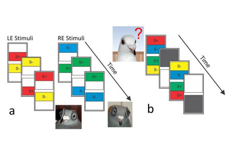

In birds each hemisphere receives visual input from the contralateral eye. Since birds have no corpus callosum, avian brains are often seen as ‘natural split brains’. But if the two hemispheres can’t interact, how do birds cope with situations, when both hemispheres are brought into conflict? Who then is in charge for a decision? If under such conditions one hemisphere completely determines the response, this is called meta-control. The aim of the current study is to test, if meta-control results from an interhemispheric conflict that would require interhemispheric interaction. If such a conflict-based interaction occurs, it should produce a delay in responding. This interaction could happen via the the commissura anterior, a small forebrain commissure that also exists in birds. To this end, biopsychologists from Bochum trained pigeons in a forced-choice color discrimination task under monocular condition such that each hemisphere was trained with a different pair of colors. Subsequently, pigeons were binocularly tested with conflicting and non-conflicting stimulus patterns. Conflicting stimuli indeed produced a delayed reaction time as expected when two divergent decisions create a conflict. Thus, pigeons indeed undergo interhemispheric conflict during meta-control even without a corpus callosum. Their hemispheres then possibly interact via the commissura anterior.

The European Union keeps millions of chicken for egg production. These animals often engage in severe feather pecking and sometimes even in cannibalistic behavior. Obviously, this causes serious welfare problems. To study the neural mechanisms of severe feather pecking, Pharmacologists from Utrecht and Wageningen universities as well as Biopsychologists from Bochum used divergent genetic lines that were selected for high or low feather pecking. These birds were analyzed for possible differences in central serotonin (5- HT) and 5-HIAA release in the limbic and prefrontal subcomponents of the caudal nidopallium by in vivo microdialysis. The results revealed that hens bred for high severe feather pecking had higher baseline levels of 5-HT in the caudal nidopallium but no differences in plasma tryptophan levels (precursor of 5-HT) and also no differences in presynaptic 5-HT storage. Thus, the data show that high aggressive encounters under industrial poultry conditions are associated with higher 5-HT releases in the prefrontal and limbic nidopallium.
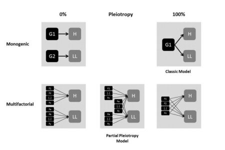

Language lateralization is one of the areas of main interest in research on human lateralization, but it is not well understood which molecular genetic factors influence this trait. Classic theories about its ontogenesis assume that it is determined by the same ontogenetic factors as handedness. In the present article, scientists from the IKN and the department of human genetics argue, that this view is contradicted by recent neuroimaging and genetics studies. Therefore, we argue that although the two traits show partial pleiotropy, there is also a substantial amount of independent ontogenetic influences for each of them. This view is supported by several recent genetic and neuroscientific studies that are reviewed in the present article.
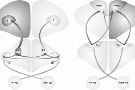

What are the neuronal and ontogenetic foundations of cerebral asymmetries? This question is deeply investigated in two avian model systems: chickens and pigeons. It is well known that the brains of these animals display physiological and anatomical left-right differences, which are related to hemispheric dominances for specific functions. In both species, asymmetrical light stimulation during embryonic development induces a dominance of the left hemisphere for visuomotor control. Nevertheless, the underlying structural asymmetries vary essentially between both species. In this recent paper, scientist from the biopsychology lab analyzed existent data on this rather puzzling phenomenon. They found evidence that although early asymmetric light stimulation is defining the dominant hemisphere for visuomotor tasks, the structural mechanisms mediating this dominance can differ between species. Furthermore, they found that environmental stimulation likely affects the balance between hemispheric-specific processing by lateralized interactions of bottom-up and top-down systems. These findings show how the interplay between environmental factors and genetically determined lateralizations can shape functional asymmetries during early development.
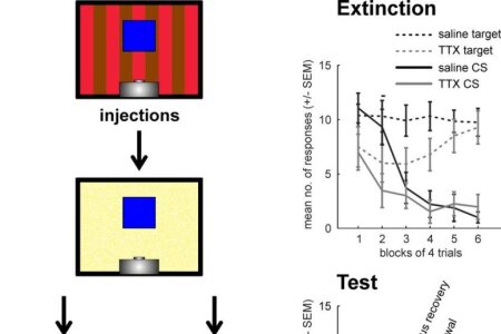

Extinction learning is an extensively investigated field of research with a general focus on fear conditioning experiments. Scientists at the Biopsychology lab shed more light on context-specific extinction learning under appetitive conditions. Here, extinction learning refers to the cessation of previously reinforced conditioned responding once reinforcement is withheld. To study the context dependency of extinction learning under appetitive conditions, we adopted and modified a within-subject ABA renewal paradigm from Robert Rescorla (Q J Exp Psychol 61: 1793) and performed pharmacological interventions to investigate fundamental neural mechanisms underlying context-dependent extinction learning. More specifically, we transiently inactivated either the nidopallium caudolaterale (NCL, functional equivalent of mammalian prefrontal cortex) or the hippocampus immediately before extinction training with tetrodotoxin. Both structures are core structures for context-specific extinction learning in fear conditioning paradigms. Using an elegantly controlled procedure, we found that both manipulations lead to non-specifically response suppression. Retrieval testing under drug-free conditions showed that subjects did successfully retrieve extinction memory in the context of acquisition but were impaired when tested in the context of extinction. Thus, the present study suggests that both NCL and hippocampus are involved in the consolidation of extinction memory, and that their contribution to extinction is context-specific.
Within a follow-up experiment we currently explore the function of NMDA-receptor in the NCL for extinction learning.


Biopsychologist Onur Güntürkün is the winner of this year's Communicator Award, conferred by the Deutsche Forschungsgemeinschaft and the Donors' Association for the Promotion of Sciences and Humanities in Germany. Professor Güntürkün, from the University of Bochum, was chosen for his exemplary approaches to communicating his research on the biological foundations for animal and human behaviour to the general public and the media. The “Communicator Award” is most important prize for science communication awarded in Germany. Established in 2000, the award is bestowed on researchers who have communicated their own scientific findings and those of their peers to a wide general audience. The prizewinners are selected by a jury comprised of science journalists and experts from the fields of public relations and communications. A total of 52 researchers working in a broad range of scientific disciplines applied or were nominated for this year's Communicator Award. After a multi-stage selection process, four of these candidates were shortlisted, with Onur Güntürkün chosen as the winner. The jury was impressed by the way in which Güntürkün combined high academic quality with a dedication to communication with the public and the media.
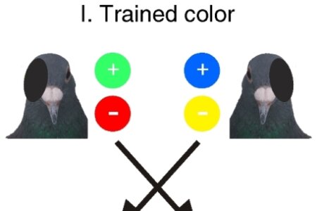

Hemispheric specialization represents a core feature of information processing in the brain of humans and other animals; however, separation of function can only be advantageous when communication systems coordinate, select and integrate information from both half brains. Scientists from the biopsychology lab have recently investigated how such systems work in pigeons and if they are influenced by envirotypic factors. Pigeons possess a lateralized visual system that is shaped by asymmetrical light stimulation during development. Comparing hemispheric-specific access to transfer information of pigeons with or without embryonic light experience demonstrates the light-modulated impact of interhemispheric communication systems. Stronger embryonic stimulation of the left hemisphere significantly enhances access to interhemispheric visual information, thereby reversing a right-hemispheric advantage that develops in the absence of embryonic light stimulation. This corroborates that environmental experiences can affect genetically determined asymmetries. This study delivers a further piece of evidence supporting the role of envirotypic factors on lateralization in the current nature vs. nurture debate.


In 2013 President Barack Obama launched the Brain Research through Advancing Innovative Neurotechnologies (BRAIN) Initiative. Comparative analyses can contribute to this effort by leading to the discovery of general principles of neural circuit design, information processing, and gene-structure-function relationships that are not apparent from studies on single species. About 30 scientists, among them two Biopsychologists from Bochum, were invited in fall 2013 to a think tank in Janelia Farm to advice the President on the BRAIN-initiative. The main goal was to come up with a strategy to extend the comparative approach to nervous system ‘maps’ comprising molecular, anatomical, and physiological data. The hope was that this research will identify which neural features are likely to generalize across species, and which are unlikely to be broadly conserved. It will also suggest causal relationships between genes, development, adult anatomy, physiology, and, ultimately, behavior. To promote this research agenda, the member of the think tank recommended that teams of investigators coalesce around specific research questions and select a set of ‘reference species’ to anchor their comparative analyses. These reference species should be chosen not just for practical advantages, but also with regard for their phylogenetic position, behavioral repertoire, well annotated genome, or other strategic reasons. This will help to form networks or consortia of researchers and centers for science, technology, and education that focus on organized data collection, distribution, and training. These activities could be supported, at least in part, through existing mechanisms at NSF, NIH, and other agencies.
Thus, our advice to the President, hoping he will read it.
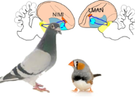

Whenever we perform a movement repeatedly, we never do it exactly the same way. There is always variability. This variability in the motor output is often viewed as noise in the system. In contrast, recent findings show that variability is more than that. Motor variability plays an important role for learning of new skills and is actively produced and regulated. Part of these findings stem from research on song birds like zebra finches. During song learning – a motor skill similar to human speech – a specialized forebrain area termed lateral magnocellular nucleus of the anterior nidopallium (LMAN) conveys variability into the motor output. Interestingly, pigeons as non-song birds possess a brain region that is possibly homologous to LMAN. This region is named nidopallium intermedium medialis pars laterale (NIML). Researchers of the Biopsychology lab together with theoretical neuroscientist from the University of Bremen have devised an experiment to test the role of NIML for behavioral variability. Pigeons learned to find hidden targets that were randomly placed at different locations on a touch screen. The researchers’ results show that their experiment induces highly variably pecking behavior. However, transient pharmacological inactivation of NIML did not result in reduction of variability. Hence, the researchers argue that in contrast to LMAN the pigeon’s NIML is not associated with behavioral variability. Together with previous findings this result suggests that LMAN’s role for variability generation in song bird is an adaptation to the special demands of song that evolved from old motor pathways of a common ancestor of recent birds. In contrast, this adaptation was not necessary in the motor system of pigeons or alternatively was lost during evolution.


We all know Christian; this rare mixture of relaxed habits, boyish charm and highest profile research. He was the boss of an Emmy Noether group in the Biopsychology lab, churning out more than 40 top papers and acquiring close to 2 million euros in the few years he was leading his small research crowd. It was clear that he wouldn’t stay for long in Bochum. Not because he didn’t liked it here (in fact, he liked it a lot) but because a young scientist with such a tremendous success rate usually becomes a professor somewhere after a short while. Now this happened. Christian became a Professor for Clinical Neurophysiology at the University Clinic for Child and Adolescent Psychiatry of the Technical University Dresden. The amount of start-up, infrastructure, and personnel he got is truly remarkable. But hey, he’s worth it! Good luck Christian!! We will miss you a lot.

On Friday the 29th of November 2013 Felix Ströckens defended his PhD thesis entitled „Evolutionary and ontogenetic processes of neuronal lateralization in vertebrates”. The title already demonstrates the wide angle of research and insight that Felix is commanding. With his experiments and analyses Felix shows that brain asymmetries have a very old past and can emerge with subtle molecular left-right differences. Most astonishingly, Felix also shows that different anatomical asymmetries can produce nearly identical lateralized behavior. In his defense Felix stood the ground, despite partly inquisitorial questions. What an impressive performance! Therefore, the committee unanimously decided to award him the title Dr. rer. nat. Congratulations Felix! We are proud of you.


Love really does change your brain. Monogamous prairie voles are known to have higher levels of oxytocin receptors than promiscuous montane voles. However, if the latter are dosed with oxytocin, they adopt the monogamous behavior of their prairie cousins. Therefore, oxytocin seems to have an important role in social bonding. But now, researchers from the University Clinic in Bonn, the University of Chengdu in China, and Biopsychologists from Bochum have performed a new experiment that suggests oxytocin stimulates the reward center in the male brain, increasing partner attractiveness and strengthening monogamy. Their study included 40 young men, all of whom had been in a relationship for at least six months and reported being passionately in love with their partners. While in a brain scanner, they either inhaled oxytocin or placebo via nasal spray while they viewed pictures of either their partners, women they knew but were not dating or women they had never met. The pictures were matched so that comparison women had been rated by independent observers as being equally attractive as the partners. In the men who were given oxytocin, the reward system (ventral tegmental area, nucleus accumbens) lit up when they saw pictures of the women they loved — but not when they looked at strangers. Some of these regions were also activated by the images of the women the men knew, but not as strongly as by the pictures of their loved ones, suggesting that it made their partners more desirable. In other words, familiarity is not enough to prompt the bonding effect of oxytocin. They must be loving couples. These results suggest that oxytocin could contribute to romantic bonds in men by enhancing their partner’s attractiveness and reward value compared with other women.
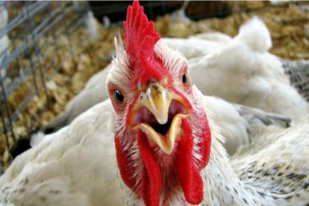

People in Germany eat more than 500 million chickens every year. The living conditions of these animals are partly abysmal. Aberrant behavior occurs often and among them severe feather pecking (SFP) cause’s major problems. SFP results in loss of feathers, skin damage and cannibalism. Now, a group of Animal Scientists from the Netherlands and Biopsychologists from Bochum studied the neurochemistry of aggression in two consecutive publications. They demonstrated that chickens with a lower tendency for SFP were less anxious and had lower levels of dopamine and serotonin turnover in somatomotor areas of the forebrain. In addition, they demonstrated that brain monoamine levels in brain areas that are involved in emotional behavior or are part of the basal ganglia-thalamopallial circuit are different in SFP-birds after a short stress period. In summary, these findings indicate that especially serotonergic neurotransmission in the dorsal thalamus, somatomotor forebrain, and striatum of hens depends on differences in behavioral feather pecking phenotype. These data show that stressful living conditions alter the serotonergic system in some individuals, leading to higher levels of anxiety and aggression.


Whether we are right- or left-handed is an important aspect of our everyday life and it comes to no surprise that handedness is the single most studied aspect of human brain asymmetries. For long it has been thought to be a monogenic trait but a single gene explaining a sufficient amount of phenotypic variance has not been identified. In the present review article, researchers from the IKN give an overview about the results of several recent studies using advanced molecular genetic techniques which suggest that a multifactorial model taking into account both multiple genetic and environmental factors, as well as their interactions, might be better suited to explain the complex processes underlying the ontogenesis of handedness. New insights into handedness genetics provided by these studies are reviewed and it is discussed, how integrating results from genetic and neuroscientific studies might help to generate more accurate models of the ontogenesis of handedness. Based on these thoughts, several new candidate gene groups whose investigation would help to further understand the complex relation of genes, the brain and handedness are discus
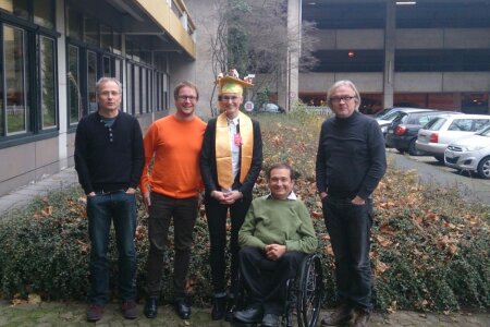

On Friday the 22nd of November Franziska Labrenz defended her PhD thesis entitled „May I capture your attention? A neurophysiological investigation into intrinsic and extrinsic determinants of visual selective attention.” Already her written thesis was impressive in the breadth of studies and methods Franziska applied to pursue her research question. In her defense she presented a marvelous talk about her main findings of the thesis, where she showed mastery in the topic of visual selective attention and important modulators of the mechanisms. It was a really impressive talk in a matchless “Franziska style”. It was a great time having you in the lab of the Emmy Noether Research group in Bochum.Congratulations Franziska!


Sex hormones have been reported to dynamically modulate the expression of implicit motives. In the present study, a team of researchers from Biopsychology, Cognitive Psychology and the Department of Psychiatry, Psychotherapy, Psychosomatics and Preventive Medicine used the Operant Motive Test (OMT) to investigate to what extent the need for affiliation, power, and achievement is affected by the menstrual cycle. In addition to measuring the strength of motive expression, the OMT also captures different forms of motive enactment. No evidence for cycle-phase related variation in overall motive scores was found. However, when different forms of motive enactment were considered, an effect of menstrual cycle was observed. The incentive-based inhibition of the power motive was significantly reduced at the time of ovulation, compared to the menstrual and to the mid-luteal phase, in naturally cycling women. In women using hormonal contraceptives, no significant changes in the form of motive enactment were evident. These results show a hormonal influence on motive-related cognitive processes.

The efficacy of executive functions depends on information processing in earlier cognitive stages. For example, initial processing of verbal stimuli in the left-hemisphere leads to more efficient response inhibition than initial processing of verbal stimuli in the right hemisphere. However, it is unclear whether this organizational principle is specific for the language system, or a general principle that also applies to other types of lateralized cognition. To answer this question, a team of IKN neuroscientists investigated the neurophysiological correlates of early attentional processes, facial expression perception and response inhibition during tachistoscopic presentation of facial ‘Go’ and ‘Nogo’ stimuli in the left and the right visual field. Participants committed fewer false alarms after Nogo-stimulus presentation in the left compared to the right visual field. This right-hemispheric asymmetry on the behavioral level was also reflected in the neurophysiological correlates of face perception, as well as in ERPs related to response inhibition. These findings show that an effect of hemispheric asymmetries in early information processing on the efficacy of higher cognitive functions can be generalized to predominantly right-hemispheric functions.

The German Research Foundation (DFG) has recently approved funding of the research unit "Extinction Learning: behavioral, neural, and clinical mechanisms", encompassing workgroups at the Universities of Bochum, Duisburg-Essen, and Marburg, for another three-year period. Extinction learning is a basic behavioral phenomenon in which an organism learns that two events which used to occur jointly have ceased doing so. Unlike acquisition, i.e. original learning, extinction is highly context-specific, which is one of the many indications that extinction is not just "unlearning" but constitutes a novel learning process distinct from original acquisition. Although extinction was first described more than 100 years ago by Russian physiologist Ivan Pavlov, many aspects of this phenomenon are still poorly understood. This is unfortunate, given that theories on the development and treatment of psychiatric disorders in part rely on extinction - e.g. psychotherapeutic interventions for phobias or psychological treatment of drug abuse. In the next three years, the research unit is expected to make significant contributions to our understanding of the neural and behavioral mechanisms of extinction learning. The scope of the projects within the unit ranges from basic research in animal models and humans to clinical applications in the treatment of phobias, enabling efficient transfer of insight gained from basic research to outpatients seeking treatment for anxiety disorders.
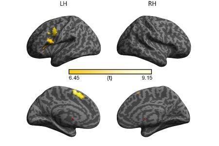

Functional hemispheric asymmetries of speech production and perception are a key feature of the human language system, but their relation to microstructural asymmetries in language-relevant white matter pathways is still poorly understood. In the present study, a team of neuroscientists from the Institute of Cognitive Neuroscience and the Bergen fMRI Group, Norway, used a combined fMRI and tract-based spatial statistics approach to investigate this question. Tract-based spatial statistics revealed several leftward asymmetric clusters in the arcuate fasciculus and uncinate fasciculus that were differentially related to functional language asymmetries. These findings suggest that white matter asymmetries may indeed be one of the factors underlying functional hemispheric asymmetries.
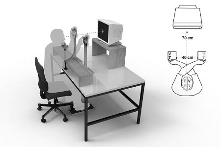

Evidence suggests that unimodal spatial processing changes across time in naturally cycling women, which is likely due to neuromodulatory effects of steroid hormones. Yet, it is unknown whether crossmodal spatial processes depend on steroid hormones as well. In the present study, a team of scientists from the Biopsychology lab, the department of Biological Psychology and Neuropsychology, University of Hamburg, and the Department of Internal and Integrative Medicine, University of Duisburg-Essen investigated this question by assessing visuo-tactile interactions in naturally cycling women, women using hormonal contraceptives and men. It was tested whether a postural effect of hand crossing on multisensory interactions in the crossmodal congruency task is modulated by women's cycle phase. It was found that visuotactile interactions changed according to cycle phase. Naturally cycling women showed a significant difference between the menstrual and the luteal phase for crossed, but not for uncrossed hands postures. The two control groups showed no test sessions effects.Regression analysis revealed a positive relation between estradiol levels and the size of crossmodal congruency effects, indicating that estradiol seems to have a neuromodulatory effect on posture processing.
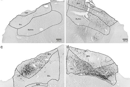

Pigeons and chickens display an asymmetry of their visual system which closely reassembles each other in behavior but not on anatomical level. While pigeons show an asymmetrically organized tectofugal system, only transient lateralizations of the thalamofugal system have been observed in chickens. By using neuronal tracing, scientists from the Biopsychology lab now analyzed if the thalamofugal system in adult, post hatch and dark incubated/monocular deprived pigeons show a comparable lateralization pattern to chickens. In all three conditions this was not the case. This indicates that visual lateralization in pigeons and chickens depends on tectofugal and thalamofugal asymmetries, respectively. Thus, in different species a highly similar pattern of behavioral asymmetries can be subserved by diverse neural systems.

Left-hemispheric language dominance is a well-known characteristic of the human language system, but the molecular mechanisms underlying this crucial feature of vocal communication are still far from being understood. In the present study, a team of scientist from the Department of Human Genetics and the Institute of Cognitive Neuroscience investigated effects of genetic variation in FOXP2 on individual language dominance. Two FOXP2 SNPs (rs2396753 and rs12533005) were found to be significantly associated with language lateralization. These results show that variation in FOXP2 may contribute to the inter-individual variability in hemispheric asymmetries for speech perception and thus provide an interesting insight into the molecular determinants of the asymmetric brain.


Previous studies from the Biopsychology lab in Bochum had shown that right head-turning preference could represent a developmental default that might influence the development of other lateralized behaviors - such as handedness - as a consequence of orienting vision towards the right side of the body. To document the role of visual experience in promoting lateralized functions, Biopsychologists from Hamburg and Bochum now assessed head-turning preference during kissing and handedness in a group of congenitally blind human adults. They found a left-side preference for head turning but a clear right-handedness in the same individuals. This unexpected asymmetric relationship has two important implications: First, the fact that absence of developmental visual experience alters the typical right head-turning pattern suggests that this motor bias is greatly affected by sensory input. Second, the study shows that visual deprivation only alters head-turn but leaves other asymmetries like handedness unaffected). The fact that experience only selectively shapes the direction of some functional asymmetries suggests that different kinds of left-right biases are organized differently. Finally it remains to be explained why blindness leads to a reversal of head-turning asymmetry. Presently only speculations can be offered. A hint to what could possibly influence such reversal comes from observation of common patterns of behavior in most blind individuals. Blind individuals tend to keep their cane in their dominant (mostly right) hand, while holding the upper right arm or the right shoulder of their guide with their left hand, so that the interlocutor is kept on the left side. Indeed, all participants of the present study used a cane daily and reported keeping it in their right hand. This particular behavior could foster selective orienting and head turning towards the left side.


The Society for the Study of Motivation (SSM) is an international, interdisciplinary organization of researchers who focus on motivation. Its mission is to encourage inquiry about all aspects of motivation from a variety of disciplines and perspectives, and to facilitate the dissemination of findings to a broad academic audience. The Society for the Study of Motivation (SSM) provides a forum for the exchange of scientific information, foster discussion of new ideas and findings on motivation among researchers, and encourages exchange and collaboration. Marlies Pinnow has now been elected as treasurer for the next period beginning in spring 2014. “It is a great honour to be a member of the SSM council and to be responsible for its financial fundament which supports all scientific, political and applied activities.” – Marlies Pinnow.


It attends to matters of general concern in research, promotes cooperation in science, advises government, parliaments and public authorities by issuing scientifically founded statements and promotes the interests of German research in relation to international science and the humanities. The Senate of the DFG has 39 members from the scientific and academic communities of which 36 are elected by the General Assembly for three-year terms. Onur Güntürkün was now elected for the period beginning with fall 2013. It is a great honor to be a member of the DFG senate. But this honor comes with the outstanding responsibility to decide on matters that deeply affect all aspects of science in Germany
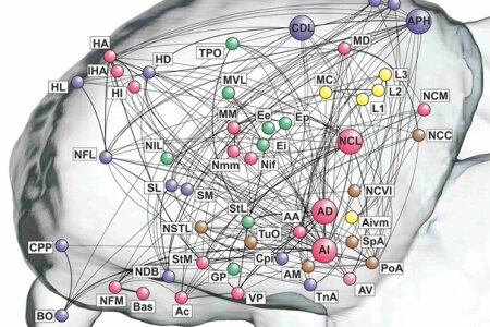

Many species of birds, including pigeons, possess demonstrable cognitive capacities, and some are even capable of cognitive feats matching those of apes. Since mammalian cortex is laminar while the avian telencephalon is nucleated, it is natural to ask whether the brains of these two cognitively capable taxa, despite their anatomical dissimilarities, might exhibit common principles of organisation on a deeper level. To find an answer to this question, a group of neuroanatomists and roboticists from Bochum, London, Auckland, Bowling Green, and Tampa came together to create the first large-scale "wiring diagram" for the forebrain of a bird. Using graph theory, they showed that, similar to mammals, the pigeon telencephalon is a modular, small-world network with a connective core of five inter-connected hub nodes that includes prefrontal-like and hippocampal structures. These regions are the most topologically central and most richly connected to the rest of the forebrain and thus central to information flow in the pigeon brain. Overall, their analysis suggests that indeed, despite the absence of cortical layers and close to 300 million years of separate evolution, the avian brain conforms to the same organisational principles as the mammalian brain on a topological level.


Handedness is the most obvious manifestation of cerebral lateralization in humans, but the molecular mechanisms that underlie its ontogenetic establishment are still poorly understood. In the present study a team of scientist from the Department of Human Genetics and the Institute of Cognitive Neuroscience performed an association study of LRRTM1 rs6733871 and a number of polymorphisms in PCSK6 and different aspects of handedness assessed with the Edinburgh handedness inventory in a sample of unrelated healthy adults (n=1113). PCSK6 rs10523972 SNP showed a significant association with a handedness category comparison and degree of handedness. These results provide further evidence for the role of PCSK6 as candidate for involvement in the biological mechanisms that underlie the establishment of handedness and support the assumption that degree of handedness, instead of direction, may be a more appropriate indicator of cerebral organization.
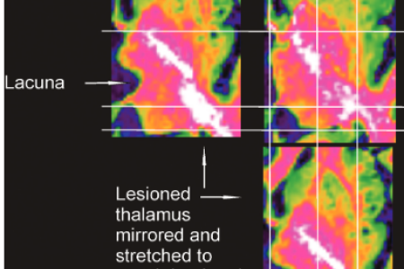

To a good extent the assessment of brain lesions in neuropsychological studies is still in its dark ages. Structural MRI-data are often grouped into arbitrary anatomical groups and are rarely analyzed with proper quantitative means that then can be correlated with assessments of cognitive decline. This problem is especially visible when it comes to thalamic lesions, where sometimes scientists lump most or at least major parts of the thalamus into a single basket. On the contrary, in animal research scientists have developed sophisticated techniques to quantify lesion loss for each and every anatomically identifiable nucleus. To overcome this problem, scientists from neuro- and biopsychology in Bochum teamed up and developed a procedure for a quantitative assessment of thalamic lesions in the chronic phase of an ischemic episode. The structural MR-images of 19 patients with ischemia in the thalamus were assessed by radiologic inspection. An independent rater allocated the damage to the each of the relevant thalamic nuclei. The assessments showed 89% accordance with the radiologic inspection. This procedure ranks the extent of the damage to thalamic nuclei and accounts for postacute rearrangement of the neural tissue. It will hopefully enable improved analyses in future studies that investigate cognitive effects of brain damages in patients.
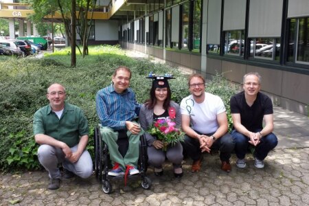

On June the 14th Vanessa Ness, member of the Emmy Noether Research group, successfully defended her PhD thesis with title „A Cognitive-neurophysiological analysis of response inhibition and action chaining“. Vanessa delivered an impressive talk about a number of high-quality studies providing an in-depth analysis of the neurophysiological mechanisms underlying complex action control. The committee was impressed by the breadth of her studies and the way Vanessa defended her results.
Congratulations Vanessa !!!

On the 5th of June 2013 Christian Beste gave his inaugural lecture on "Cognitive Neurophysiology of Action Control: Between Basic and Applied Science". It was a fantastic tour de force of neuroscientific experiments that proceeded all the way from cells to patients. With this lecture the process of habilitation is now officially completed.
Congratulations Christian!


The Marsilius Lectures are activities for the academic community of all disciplines as well as for the broader public. Once a semester, the Marsilius Kolleg invites a well-known scientist to speak about a topic that calls for bridging the gaps between scholarly cultures. In recognition of their achievements in the dialogue between scholarly cultures, the speakers of the Marsilius Lecture are decorated with the Marsilius Medal. This May it was Onur’s turn. In a stunningly baroque and huge lecture hall he spoke about “Die Evolution des Denkens”. Afterwards he was awarded with the Marsilius Medal.

The excitability of our cortex varies between individual and over time. Does this variability affect learning speed and even behavioral variability? Indeed, facilitation of motor cortical excitability has been shown to be positively correlated with improvements in performance in simple motor tasks. Thus cortical excitability may tentatively be considered as a marker of learning and use-dependent plasticity. Previous studies focused on changes in cortical excitability brought about by learning processes, however, the relation between native levels of cortical excitability on the one hand and brain activation and behavioral parameters on the other is as yet unknown. A team of Neurologists and Biopsychologists from Bochum now investigated the role of differential native motor cortical excitability for learning a motor sequencing task. The motor task required the participants to reproduce and improvise over a pre-learned piano sequence. Over both task conditions, participants with low cortical excitability showed significantly higher BOLD activation in task-relevant brain regions than participants with high cortical excitability. In contrast, both groups did not exhibit differences in learned performance and improvisation level. Moreover, cortical excitability did practically not change after learning and training in either group. The present data suggest that the native, unmanipulated level of cortical excitability is related to brain activation intensity, but not to performance quality. Possibly, the subjects with the lower cortical excitability possibly only showed a higher BOLD signal intensity to compensate for their lower level of excitation.
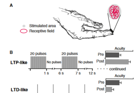

It sounds like an old dream is becoming true. Recent work suggests that intensive training may not be necessary for skill learning. Skills can be effectively acquired by a complementary approach in which the learning occurs in response to mere exposure to repetitive sensory stimulation. Such training-independent sensory learning induces lasting changes in perception and goal directed behaviour in humans, without any explicit task training. We suggest that the effectiveness of this form of learning in different sensory domains stems from the fact that the stimulation protocols used are optimized to alter synaptic transmission and efficacy. While this approach directly links behavioural research in humans with studies on cellular plasticity, other approaches show that learning can occur even in the absence of an actual stimulus. These include learning through imagery or feedback-induced cortical activation, resulting in learning without task training. All these approaches challenge our understanding of the mechanisms that mediate learning. Apparently, humans can learn under conditions thought to be impossible a few years ago. Although the underlying mechanisms are far from being understood, training-independent sensory learning opens novel possibilities for applications aimed at augmenting human cognition.
Beste, C., Dinse, H.R. (2013). Learning without training. Curr Biol, 23, 489-499.
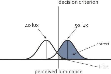

A vast body of data supports the notion that animals, including humans, perform statistically optimally in a wide range of tasks, supporting the claim that evolution has shaped the nervous system of organisms in a way that yields maximally adaptive behavior. Optimality is frequently assessed by comparing behavioral output to benchmarks computed via methods derived from statistical decision theory. Such methods have also been used to assess the reliability of sensory neural signals, and have even been invoked as accounts of neural processing.Scientists from the Biopsychology lab have explicitly tested the claim of optimality with pigeons performing a perceptual choice task with varying reward probabilities. Surprisingly, the pattern of choices observed was opposite to that expected under an optimization account. This finding poses important constraints on the class of algorithms useful for modeling adaptive choice behavior. Furthermore, the algorithm which fit the data best was learning exclusively on correct trials ending in reward, as opposed to algorithms learning from both reward and errors. Put simply, the pigeons seem to learn from reward, but not from errors - at least when performing a psychophysical task.
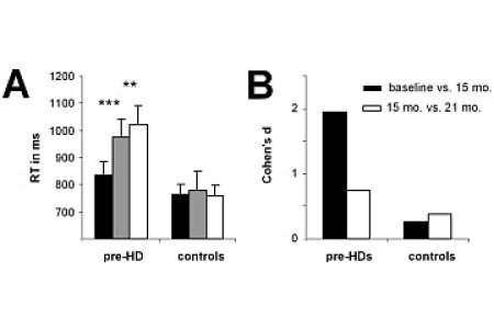

In several neurodegenerative diseases, like Huntington's disease (HD), treatments are still lacking. To determine whether a treatment is effective, sensitive disease progression biomarkers are especially needed for the premanifest phase, since this allows the evaluation of neuroprotective treatments preventing, or delaying disease manifestation. On the basis of a longitudinal study we present a biomarker that was derived by integrating behavioural and neurophysiological data reflecting cognitive processes of action control. The measure identified is sensitive enough to track disease progression over a period of only 6 month. Changes tracked were predictive for a number of clinically relevant parameters and the sensitivity of the measure was higher than that of currently used parameters to track prodromal disease progression. The study provides a biomarker, which could change practice of progression diagnostics in a major basal ganglia disease and which may help to evaluate potential neuroprotective treatments in future clinical trials.
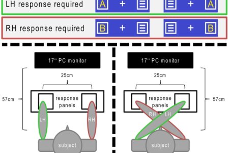

It is well-kown that sensory information influences the way we execute motor responses. However, less is known about if and how sensory and motor information are integrated in the subsequent process of response evaluation. We used a modified Simon Task to investigate how these streams of information are integrated in response evaluation processes, applying an in-depth neurophysiological analysis of event-related potentials (ERPs), time-frequency decomposition and sLORETA. The results show that response evaluation processes are differentially modulated by afferent proprioceptive information and efference copies. While the influence of proprioceptive information is mediated via oscillations in different frequency bands, efference copy based information about the motor execution is specifically mediated via oscillations in the theta frequency band. Stages of visual perception and attention were not modulated by the interaction of proprioception and motor efference copies. Brain areas modulated by the interactive effects of proprioceptive and efference copy based information included the middle frontal gyrus and the supplementary motor area (SMA), suggesting that these areas integrate sensory information for the purpose of response evaluation. The results show how motor response evaluation processes are modulated by information about both the execution and the location of a response.


Can you trust this man? What sounds like an everyday question, is indeed a classic problem of human evolution, especially as seen from a female perspective. According to evolutionary theories, women should have different levels of trust dependent on their actual menstrual cycle. This prediction was tested by Biological Psychologists and Psychiatrists from Bochum University. They tested how trusting behavior varies in naturally cycling women, as a function of sex and attractiveness of players in a trust game, at three distinct phases of the menstrual cycle. Women acted more cautiously in an investment game at the preovulatory phase, compared to the menstrual and the mid-luteal phase. Reduced willingness to trust in strangers was particularly expressed toward male players at this time. The increase of estradiol levels from menses to the preovulatory phase was negatively correlated with trust in attractive male other players, whereas the increase of progesterone levels from menses to the mid-luteal phase was positively associated with trust in unattractive female other players. Thus, the results emphasize the impact of the menstrual cycle on interpersonal trust, although the exact mode of hormonal action needs to be further investigated.
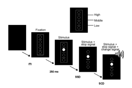

Our daily life is characterized by multiple response options that need to be cascaded in order to avoid overstrain of restricted response selection resources. While response selection and goal activation in action cascading are likely driven by a process varying from serial to parallel processing, little is known about the underlying neural mechanisms that may underlie interindividual differences in these modes of response selection. To investigate these mechanisms, we used a stop–change paradigm for the recording of event-related potentials and standardized low resolution brain electromagnetic tomography source localizations in healthy subjects. Systematically varying the stimulus onset asynchrony (the temporal spacing of “stop” and “change” signals), we applied mathematical constraints to classify subjects in more parallel or more serial goal activators during action cascading. On that basis, the electrophysiological data show that processes linking stimulus processing and response execution, but not attentional processes, underlie interindividual differences in either serial or parallel response selection modes during action cascading. On a systems level, these processes were mediated via a distributed fronto-parietal network, including the anterior cingulate cortex (Brodman area 32, BA32) and the temporo-parietal junction (BA40). There was a linear relation between the individual degree of overlap in activated task goals and electrophysiological processes.
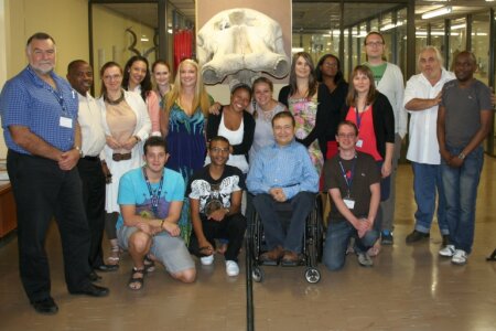

During the last week, the "First South-Africa-German Summer school on comparative psychology" took place in one of the most exciting cities of the southern hemisphere: Johannesburg. Supported by the National Research Foundation of South Africa and the BMBF, students, young academics and senior scientist from the biopsychology lab and the department of anatomical science of the University of Witwatersrand had the opportunity to discuss their projects, plan new experiments and intensify South African -- German cooperation in the field of neuroscience.From damaraland mole rat courtship behavior over elephant hippocampus research to cetacean neuroanatomy and many other research areas the school covered a wide range of fascinating topics within the comparative psychology of unusual African model species.Receiving overwhelming positive feedback from all participants we agree that this summer school was a great experience and we are looking forward to further cooperations and activities with our old and new friends from South Africa.
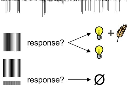

It is important to know which events in our environment are predictive of reward or punishment. For example, knowing that the sound of the doorbell signals postal delivery while the sound of the fire alarm requires leaving the house both inform us about the present state of our world as well as about appropriate actions to take. This ability to decide on a course of action given the current state of the world is implemented in a widely distributed neural network, and one suspected hot spot for decision making is the pigeon nidopallium caudolaterale (NCL).
In the present study, neurophysiologists from the biopsychology lab recorded single-unit activity in this forebrain area while animals were working on a visual discrimination task where responding or non-responding could lead to reward, punishment, or be inconsequential. A sophisticated analysis of the NCL neurons’ activation patterns revealed that these neurons represent information about the reward value of specific stimuli, instrumental actions as well as action outcomes, and therefore provide signals useful for adaptive behavior in dynamically changing environments.
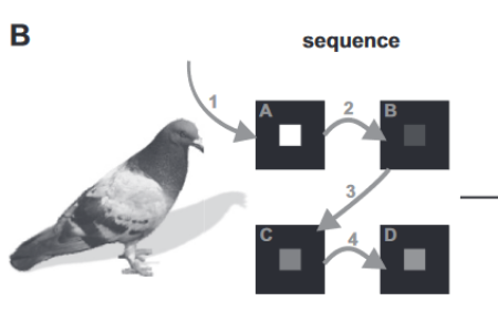

Most of our everyday activities consist of action sequences as for instance brushing your teeth, shoe lacing or driving a car. The pigeon is a classic animal model for studying the fundaments of sequence learning and execution. Yet, little is known about the neural basis mediating sequences in pigeons. In a recent study, scientists from the Biopsychology department in collaboration with the Mercator Research Group "Structure and Memory" identified two forebrain structures in the pigeon brain that play a pivotal role in mediating a memorized sequence.
Researchers of the Biopsychology department already showed in a previous study (Helduser & Güntürkün, 2012) that the nidopallium caudolaterale (NCL) and the nidopallium intermedium medilalis pars laterale (NIML) are associated with sequence execution. However, in the previous study the pigeons' behavior was cue guided. To clarify the role of both structures for memorized sequences, the authors of the recent study applied a purely memory based task. Pigeons learned to peck a four item-sequence on a touch screen. Pharmacological inactivation both of NCL and NIML impaired sequence execution confirming previous results that NCL and NIML store and process sequences in parallel. In addition, a special role of NCL for sequence initiation was revealed.
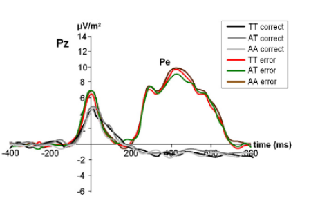

Action control mechanisms are modulated by a number of factors related to neurobiology and affective states like anxiety. However, the precise interrelation of these factors is widely unknown. The neuropeptide S (NPS) system has been suggested to contribute to the pathogenesis of anxiety. In order to further characterize the cognitive-neurophysiological relevance of neuropeptide S in the etiology of anxiety, the influence of a functional neuropeptide S receptor gene (NPSR1) variant on response inhibition and error monitoring was investigated under consideration of the dimensional phenotype of anxiety sensitivity (AS). In a sample of healthy probands, event-related potential (ERP) measurement using a modified Flanker task was applied allowing for a distinct neurophysiological examination of processes related to response inhibition and error monitoring. All subjects were genotyped for the functional NPSR1 A/T (Asn107Ile) variant (rs324981) and characterized for anxiety sensitivity using the Anxiety Sensitivity Index (ASI). Carriers of the NPSR1 T allele displayed intensified response inhibition and error monitoring, which was in both cases paralleled by the behavioral data. Furthermore, anxiety sensitivity was found to be higher in NPSR1 T allele carriers and to correlate with Nogo-P3 and Ne/ERN. A mediation analysis revealed the ERN to mediate the effect between NPSR1 genotype and anxiety sensitivity. In summary, the more active NPSR1 T allele may confer enhanced response inhibition and increased error monitoring and might drive particularly error monitoring as a neurophysiological endophenotype of anxiety as reflected by increased anxiety sensitivity. The study is among the first which is able to directly integrate mechanisms underlying action control with factors of 'anxiety' and 'genetics' into a common framework.

Language lateralization is a key feature of human brain organization, but the molecular mechanisms underlying its ontogenesis are still poorly understood. In the present study a team of scientist from the Department of Human Genetics and the Institute of Cognitive Neuroscience investigated whether variation in schizophrenia-related genes modulates individual lateralization patterns. For the first time, a significant association of genetic variation in the cholecystokinin-A receptor (CCKAR) and language lateralization was found. Individuals carrying the schizophrenia risk allele C of this polymorphism showed a marked reduction of the typical left-hemispheric dominance for language processing. Since the cholecystokinin A receptor is involved in dopamine release in the central nervous system, these findings suggest that genetic variation in this receptor may modulate language lateralization due to its impact on dopaminergic pathways.


On the 18th of December 2012 Denise Manahan-Vaughan stepped down from as speaker of the Research Department of Neuroscience. During her term of office, the Research Department was founded and an excellent infrastructure of jointly used research facilities was established. Now as her term was over, Onur Güntürkün was elected anonymously. The elected Vice speakers are Stefan Herlitze and Ulf Eysel. The following term has its own challenges: Money is much more limited than it was before and major collaborative research grants await renewal. The picture shows Denise Manahan-Vaughan and Onur Güntürkün after the elections.
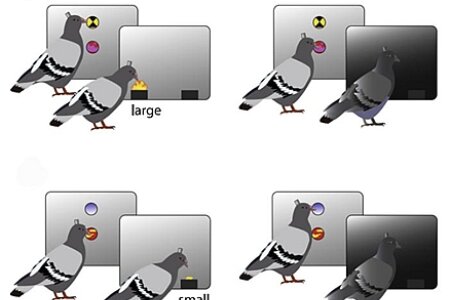

We quickly learn that some of our actions result in reward. But reward comes in different magnitudes and we should be able to repeat those behaviors that are associated with the highest amount of reinforcement. In a new study, Biopsychologists from Boston, Oxford, and Bochum investigated the contribution of striatal dopamine receptors (D1) on the learning of reward-magnitude. To this end, pigeons were trained on a discrimination-task with two pairs of stimuli; correct discrimination resulted in a large reward in one pair of stimuli and in a small reward in the other pair. Acquisition of the discrimination-task was accompanied by intracranial injections to the medial striatum, either of a dopamine-antagonist (Sch23390) or of vehicle. In the control-condition the rate of learning was modulated by the magnitude of the reward. Following injections of D1 antagonist this effect vanished even though the ability to discriminate between the rewards was unaffected. Interestingly, the mean rate of learning was indistinguishable between the control and antagonist conditions. Consequently, it appears that not learning per se but the effect of reward-magnitude on learning is mediated through D1 receptors in the striatum. The intriguing possibility is this: It is conceivable that the injections of the dopamine-antagonist caused a shift in learning strategy. In the control-condition animals possibly relied on positive feedback and thus their learning was affected by the magnitude of the contingent reward; in the antagonist-condition, however, learning might have relied on negative feedback and was thus insensitive to reward-magnitude.

Onur Güntürkün accepted the invitation to become a member of the Editorial Board of Brain, Behavior and Evolution (Karger). Birds are meanwhile a key taxonomic group to understand the evolution of brain and behavior in vertebrates. And in this endeavor, the analysis of cognition gains importance. These were the reasons why Georg Striedter, the Editor-in-Chief of Brain, Behavior and Evolution invited Onur Güntürkün to come onboard to help steering this journal.
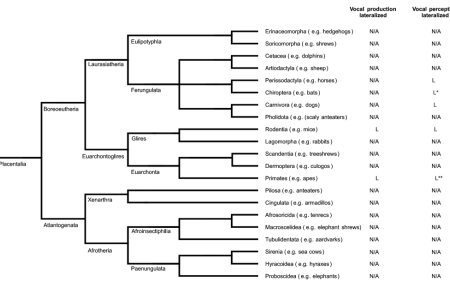

Lateralization of conspecific vocalization has been observed in several vertebrate species but a systematic integration of these results across orders was lacking so far. Biopsychologists from Bochum now used cladographic comparisons to identify those vertebrate orders in which evidence for or against lateralization of conspecific vocalization has been reported, and those orders in which further research is necessary. The analysis showed that there is evidence for lateralization of conspecific vocalization in several mammalian orders (e.g. Primates) and also evidence for lateralization of conspecific vocalization in some avian species (e.g. within the Passeriformes order). While especially the primate data suggest that human language lateralization could have resulted from an inherited dominance of the left hemisphere for those neural properties of language that are shared with the sensory or motor aspects of vocalizations in other vertebrate species, it becomes clear that this conclusion presently is supported by only sparse empirical evidence. The majority of vertebrate orders, especially among non-amniotes, still need to be explored.
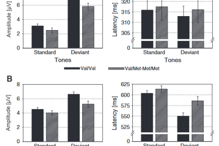

Aging affects the ability to focus attention on a given task and to ignore distractors. These functions subserve response control processes for which fronto-striatal networks have been shown to play an important role. Processing efficacy in these networks crucially depends upon BDNF. We investigated how cognitive subprocesses constituting a cycle of distraction, orientation and refocusing of attention are affected by the functional BDNF Val66Met polymorphism using event-related potentials (ERPs) in healthy elderly. We found that the Val/Val genotype confers a disadvantage to its carriers. This disadvantage was partly compensated by intensified attentional shifting mechanisms. It could be based on response selection processes being more vulnerable against interference from distractors in this genotype group. Processes reflecting transient sensory memory processes, or the re-orientation of attention were not affected by the BDNF Val66Met polymorphism, suggesting a higher importance of BDNF for mechanisms related to response control, than stimulus processing. The results add on recent literature showing that the Met allele confers some benefit to its carriers. We suggest an account for unifying different results of BDNF Val66Met association studies in executive functions, based on the role of BDNF in fronto-striatal circuits.
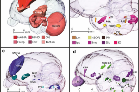

Pigeons are classic animal models for learning, memory, and cognition. The majority of the current understanding about avian neurobiology outside of the domain of the song system has been established using pigeons. Since MRI represents an increasingly relevant tool for comparative neuroscience, a 3-dimensional MRI-based atlas of the pigeon brain becomes essential. Using multiple imaging protocols, a team from the Biopsychology in Bochum and the Bioimaging Centre in Antwerp delineated diverse ascending sensory and descending motor systems as well as the hippocampal formation. The resulting pigeon brain atlas can easily be used to determine the stereotactic location of identified neural structures at any angle of the head. The atlas is freely available for the scientific community. Just click on the button with the scan of the pigeon head to the left of this text.


Running a big university lab is like continuously juggling a dozen balls. Every moment multiple tasks have to performed, projects that unfold in timescales of days to years have to be monitored, kilograms of papers have to reviewed and produced, and, finally, literature has to be read, understood and integrated into an ever-growing tapestry of knowledge. At the same time, competition is tough and reputation that has been built in decades can be destroyed in a day. There simply is no time to deeply think about issues that need weeks to grow in your mind.
Therefore Onur Güntürkün accepted the invitation to apply for a stay in the Wissenschaftskolleg zu Berlin (WiKo). There, in Berlin Grunewald, scientists can move into apartments within a classic villa and just think deeply for up to one year, while WiKo pays their salary to the home university for teaching replacement. Now, WiKo accepted him a fellow. The plan is not to stay for full 12 months but to take a stay from October 2014 to April 2015.
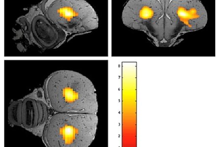

Functional MRI (fMRI) is widespread in humans but still rare in animal research. If being applied, nearly always anaesthetized animals are used. Only a few articles present results obtained in awake rodents. With advent of the "cognitive bird revolution", awake bird fMRI is increasingly relevant. Bioimaging experts from Antwerp and Biopsychologists from Bochum now conducted the first awake bird imaging study with highly habituated and head fixed pigeons. Both traditional fMRI and resting state (rsfMRI) were applied. In addition, this is the first time functional connectivity measurements were performed in a non-mammalian species. Since the visual system of pigeons is a well-known model for brain asymmetry, the focus of the study was on the neural substrate of the visual system. For fMRI a visual stimulus was used and functional connectivity measurements were done with the entopallium as a seed region. Interestingly in awake pigeons the left E was significantly functionally connected to the right E. Moreover connectivity maps for a seed region in both hemispheres resulted in a stronger bilateral connectivity starting from left E. These results could be used as a starting point for further imaging studies in awake birds and also provide a new window into the analysis of hemispheric dominance in the pigeon.


In humans, interpersonal romantic attraction and the subsequent development of monogamous pair-bonds is substantially predicted by influential impressions formed during first encounters. The prosocial neuropeptide oxytocin (OXT) has been identified as a key facilitator of both interpersonal attraction and the formation of parental attachment. However, whether OXT contributes to the maintenance of monogamous bonds after they have been formed is unclear. Psychiatrists from Bonn, Biopsychologists from Bochum and Neuroscientists from Chengdu now provide the first behavioral evidence that the intranasal administration of OXT stimulates men in a monogamous relationship, but not single ones, to keep a much greater distance (10 –15 cm) between themselves and an attractive woman during a first encounter. They further confirmed this using a photograph-based approach/avoidance task that showed again that OXT only stimulated men in a monogamous relationship to approach pictures of attractive women more slowly. Importantly, these changes cannot be attributed to OXT altering the attitude of monogamous men toward attractive women or their judgments of and arousal by pictures of them. Together, these results suggest that where OXT release is stimulated during a monogamous relationship, it may additionally promote its maintenance by making men avoid signaling romantic interest to other women through close-approach behavior during social encounters. In this way, OXT may help to promote fidelity within monogamous human relationships.


On the 6th of December 2012 The Senate of the Deutsche Forschungsgemeinschaft decided to award Onur Güntürkün with the Gottfried Wilhelm Leibniz Award. With 2.5 million Euro that can freely be spent within 7 years, the Leibniz award is the highest European award of its kind. The DFG calls it "freedom like in fairy tales".
It feels like that.

Sascha Helduser successfully graduated on the 5th of December 2012 and was awarded with a PhD of Neuroscience. Sascha was able to achieve major insights into the neural basis of sequence learning and sequential behavior in pigeons. Given that many insights into the behavioral fundaments of sequencing were gained by studying pigeons, it was amazing that practically nothing was known about the brain systems that mediate this type of behavior. By training pigeons in various complex behavioral studies and subsequently blocking the activity of different brain areas, Sascha was able reveal that two brain structures were especially relevant. One was the NCL, the bird equivalent of the prefrontal cortex. The other was the pallial component of a "cortico"-basal ganglia-thalamus loop. The name of this structure in song birds is LMAN (please have a look at his t-shirt!). Both of these two structures contribute to sequential behavior, albeit slightly differently. In his final experiment Sascha was even able to increase the success of sequence execution by microstimulating the NCL shortly before sequence onset. The committee was truly impressed with Sascha's achievements and decided to award him the doctorate title with the grade magna cum laude.

On Tuesday the 27th Stefanie Schulz received her PhD (Dr. rer. nat.) for her work on neurogenetic foundations of attentional processes. In her studies she examined the role of various single nucleotide polymorphisms for attentional processes within the framework of the attention network test. Already her written thesis was impressive. In a marvelous defense of her work she impressed the committee with her in-depth knowledge on attentional processes and neurobiological foundations thereof. It was a great having you in the Biopsychology, Motivational Psychology and Emmy Noether Group team.


Since several years Tagrid is giving the Neurophysiology lecture on behalf of the chair of Biopsychology. Since then our students start with a comprehensive and perfectly presented knowledge about neuronal functions into their studies. Now the outstanding pedagogical skills of Tagrid were awarded by the highest possible institution. A consortium consisting of the German Philological Society and several foundations selected just ten out of 3,500 nominated teachers for this high honor; and Tagrid was one of these few selected ones. The key issue is that teachers can't apply; their former students nominate and vote for them! Tagrid received the award for her fantastic commitment to teaching, her approachability, and her dedication to transmit the full complexity of the content in the best possible way. Tagrid shows up at dawn in the slaughterhouse to snatch a pig brain for her class and she is faced with children from a societal background as remote from academia as you can imagine. But she is standing the ground and delivers her message that science is the key to the future.


Complex regional pain syndrome (CRPS1) is a chronic progressive disease characterized by severe pain without demonstrable nerve lesion. It often affects an arm or a leg and may spread to another part of the body. In a previous study biopsychologists and neurologists from Bochum had shown that CRPS-patients have a remarkable shift of the visual subjective body midline, a correlate of the egocentric reference frame, towards the affected side (Reinersmann et al., 2010). However, this first study fell short of showing if this asymmetrical change of the egocentric computation was specific for CRPS. Now, the same team is able to report that this asymmetric subjective shift towards the left side is specific for CRPS1-patients with right sided pain and does not occur in controls or in patients with other types of chronic pain. Thus, right-affected CRPS1-patients display an asymmetrical attentional spatial focus that is thought to result from unilateral CRPS pain inducing a somatosensory imbalance, with pain providing ''exaggerated'' input from the affected side. This then could distort visuospatial perception, including perceptual representation of one's own body. This study shows that CRPS1 is a syndrome of sensory-motor-autonomic dysfunctions that goes along with neural changes that control lateralized attentional systems.
The editors of the journal thought this discovery to be of great importance and associated it with an invited commentary (Greenspan, 2012).


In a previous study biopsychologists from Bochum and Nürnberg had demonstrated that bottlenose dolphins can discriminate visual stimuli according to their numerosity (the number of objects displayed in pictures). But are dolphins able to use an abstract numerical category based on "few" vs. "many" when discriminating stimuli according to the number of their constituent patterns? And how do they mentally represent numerosity? To this end, a team of Biopsychologists from Bochum, Mallorca and Nürnberg trained Blue, an adult bottlenose dolphin, to discriminate between two simultaneously presented stimuli which varied in the number of elements they contained. After initial training, several confounding parameters were excluded to show that discrimination performance indeed depended on numerosity. Subsequently, the animal was tested with new stimuli of intermediate as well as higher numbers of elements. Once discrimination had been achieved, a reversal-training on a subset of stimuli was initiated. Afterward, the subject generalized the reversal successful to new and unreinforced stimuli. These results reveal two main findings: firstly, after reversal learning, Blue generalized the reversal successful to new and unreinforced stimuli. Thus, the results suggest that dolphins are able to learn and use a numerical category that is based on abstract qualities of "few" vs. "many." In addition, the occasional errors of Blue strongly suggest a magnitude and a distance effect. Thus, coding of numerical information in dolphins might follow logarithmic scaling as postulated by the Weber-Fechner law.
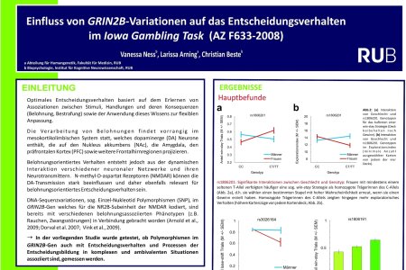

Vanessa Neß participated in this year's "FoRUM" conference hosted by the medical faculty of the RUB. For her poster presentation summarizing a cooperation project with the human genetics group, funded by FoRUM (Forschungsförderung Ruhr-Universität Bochum Medizinischen Fakultät), she won a poster prize in her category endowed with a prize money of 500€.The dopaminergic system is known to modulate decision-making. With N-methyl-D-aspartate receptors (NMDARs) strongly influence dopaminergic functioning, it is conceivable that the glutamatergic system is also involved in decision-making. The GRIN2B gene codes for the NMDAR subunit 2B determining receptor affinity. Out of 15 polymorphisms of the GRIN2B gene, two showed a strong association with strategic aspects of IGT performance. The results suggest that healthy individuals with certain GRIN2B variations respond differently to ambiguous conditions, possibly by altered perception of wins and losses. These findings underline the necessity to integrate the glutamatergic system when examining decision making processes.
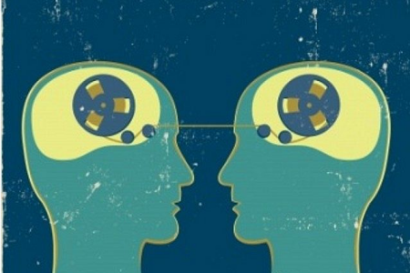

Humans incur considerable costs to punish unfairness directed towards themselves or others. Recent studies using repetitive transcranial magnetic stimulation (rTMS) suggest that the right dorsolateral prefrontal cortex (DLPFC) is causally involved in such strategic decisions. Presently, two partly divergent hypotheses are discussed, suggesting either that the right DLPFC is necessary to control selfish motives by implementing culturally transmitted social norms, or is involved in suppressing emotion-driven prepotent responses to perceived unfairness. Accordingly, a team of psychiatrists and biopsychologists from Bochum studied the role of the DLPFC in costly (i.e. third party) punishment by applying rTMS to the left and right DLPFC before playing a Dictator Game with the option to punish observed unfair behavior (DG-P). Costly punishment increased, albeit nonsignificantly, upon disruption of the right – but not the left – DLPFC as compared to sham stimulation. However, empathy emerged as a highly significant moderator variable of the effect of rTMS over the right, but not left, DLPFC, suggesting that the right DLPFC is involved in controlling prepotent emotional responses to observed unfairness, depending on individual differences in empathy.
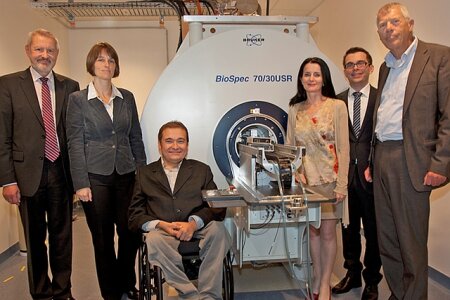

On the 22nd of October 2012 the new Bruker 7 Tesla animal MRT-scanner facility was opened with a nice ceremony. The system was awarded by the Mercator Foundation to the Mercator Research Group „Structure of Memory“. The facility includes, besides the scanner, a surgery and a testing room as well as animal housing facilities for mice, rats, and pigeons. In addition, a physicist from Caltech was hired to run the system. The first pictures taken were from mandarins and kiwis, but meanwhile mice, rat, and pigeon brains were also successfully analyzed. The goal for the Biopsychology lab will be to establish awake pigeon fMRI.


Recently, a dispute on the presence or absence of asymmetry of the avian magnetic compass was discussed in Nature (Nature, 2011, 471: E12-13). A new study from biologists in Frankfurt and biopsychologists in Bochum could clarify some of the contradictions. They now revealed that the lateralized magnetic compass of the migratory European robin is not asymmetrically organized from the ontogenetic beginning, but develops as the birds grow older. During first migration in autumn, juvenile robins can orient by their magnetic compass with both eyes. In the following spring, however, the magnetic compass is already lateralized towards the right eye/left hemisphere, but this lateralization is still flexible: it could be removed by covering the right eye for 6 h. During the following autumn migration, the lateralization becomes more strongly fixed, with a 6 h occlusion of the right eye no longer having an effect. This change from a bilateral to a lateralized magnetic compass appears to be a maturation process, the first such case known so far in birds. Because both eyes mediate identical information about the geomagnetic field, brain asymmetry for the magnetic compass could increase efficiency by setting the other hemisphere free for other processes.

Glutamatergic neural transmission is involved in both neural plasticity and neurodegeneration. This combination of roles could result in ambivalent effects in which excitotoxic neurodegeneration augments neural plasticity in parallel. Neural plasticity can be induced by exposure-based learning (EBL) that resembles timing properties of long-term potentiation (LTP) protocols (i.e., LTP-like learning). Even though it has not been demonstrated so far in animal models that perceptual effects of such stimulation protocols are mediated by typical LTP mechanisms, it has been shown that exposure-based learning exerts strong effects on cognitive brain functioning and is modulated by glutamatergic neural transmission. We reveal that exposure-based perceptual learning is more efficient in a human model of excitotoxic neurodegeneration than in healthy participants. Pre-manifest Huntington's disease gene mutation carriers showed faster increases in perceptual sensitivities than controls. This in turn changed attentional processing in extrastriate visual areas objectified using electroencephalogram data. The emergence of faster learning correlated positively with genetic disease load. Our results confirm an ambivalent action of increased glutamatergic transmission, implying that the process of excitotoxic neurodegeneration is associated with enhanced perceptual learning, which can be used to improve attentional and behavioral control via the alteration of perceptual sensitivities.
“A thought-provoking new study has found that symptom-free carriers of the neurodegenerative Huntington’s disease present a dramatic two-fold acceleration in perceptual learning.” (Cardoso-Leite et al., 2012).
“These results are spectacular in the face of the massive literature showing subtle cognitive impairments in pre-HD patients.” (Cardoso-Leite et al., 2012).
“Adapting the exposure-based learning procedure pioneered in the present study to animal models in future research could be a productive way to characterize the respective mechanisms subtending enhanced learning and excitotoxicity in Huntington’s disease.” (Cardoso-Leite et al., 2012).
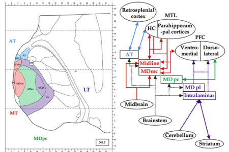

The functional role of the mediodorsal thalamic nucleus (MD) and its cortical network for recognition and memory recall is discussed controversially. Neuropsychologists and Biopsychologists from Bochum now decided to take a fresh approach: They studied patients with focal thalamic lesions to examine the functions of the MD, the intralaminar nuclei and the midline nuclei in memory processing. In addition, a newly designed pair association task was used, which allowed the assessment of recognition and cued recall performance. Most importantly, volume loss in thalamic nuclei was estimated with a technique that had been invented in animal research but was used in this study for the first time on humans. These quantitative anatomical data could then be used as a predictor for alterations in memory performance. The results reveal that reduced recall of picture pairs and increased response times during recognition followed by cued recall covaried with the volume loss in the parvocellular MD. This pattern suggests a role of this thalamic region in recall and thus recollection, which does not fit into the classic framework that is in use in today's Neuropsychology. The functional specialization of the parvocellular MD accords with its connectivity to the dorsolateral PFC, highlighting the role of this thalamocortical network in explicit memory.


The German Psychological Society decided to award Sebastian Ocklenburg this year’s Heinz-Heckhausen Young Investigator Award for his outstanding dissertation. During the laudatio, Prof. Oliver Wolf outlined that the committee of the German Psychological Society was deeply impressed by the quality and the quantity of publications that constitute Sebastian’s thesis. Oliver Wolf then went on to say that the breadth of the studies on cerebral asymmetries conducted by Sebastian start to lay the fundaments of a theory of brain asymmetry. The Heinz-Heckhausen Young Investigator Award is only given once in two years during the Congress of the German Psychological Society.
CONGRATULATIONS SEBASTIAN !!!


Die "First South African-German summer school on comparative psychology" ist ein professionelles Fortbildungsprogramm innerhalb der vergleichenden Psychologie, einer neu aufkommenden Fachrichtung innerhalb der Geisteswissenschaften, die sich mit den evolutionären Hintergründen der menschlichen Kognition beschäftigt.
Die Summer School wird unter der Leitung von Dr. Nina Patzke und Prof. Paul Manger in Zusammenarbeit mit der Arbeitseinheit Biopsychologie, an der Universität von Witwatersrand, Johannesburg, Südafrika im Februar 2013 stattfinden und aus einer Mischung von wissenschaftlichen Vorträgen, Projektdiskussionen und dem Training von praktischen Forschungstätigkeiten bestehen.
Die Summer School findet vom 18. bis zum 22. Februar 2013 an der Universität von Witwatersrand, Johannesburg, Südafrika statt. Zielgruppe sind junge, südafrikanische Wissenschaftler innerhalb des Master- oder Promotionsstudiums, die sich für den Bereich der vergleichenden Psychologie interessieren. Bewerben können sich Wissenschaftler aus dem Feld der Geisteswissenschaften, Biologie, Psychologie oder Medizin. Wir möchten besonders junge Wissenschaftlerinnen ansprechen, da die Summer School eine spezielle Vortragsreihe mit dem Titel "women in science" (siehe Programm) enthält, in dem zwei erfolgreiche Wissenschaftlerinnen aus Deutschland und Südafrika über Ihren Werdegang, sowie Chancen und Karrieremöglichkeiten für Frauen in der Wissenschaft berichten. Es wird eine möglichst kleine Gruppengröße angestrebt um den Austausch zwischen Teilnehmern und den vortragenden Wissenschaftlern zu erleichtern und individuelle Beratungsgespräche zu ermöglichen. Daher ist die Anzahl der Teilnehmer auf 10 Bewerber begrenzt.


Attentional mechanisms are a crucial prerequisite to organize behavior. Most situations may be characterized by a ‘competition’ between salient, but irrelevant stimuli and less salient, relevant stimuli. In such situations top-down and bottom-up mechanisms interact with each other. In the present fMRI study, we examined how interindividual differences in resolving situations of perceptual conflict are reflected in brain networks mediating attentional selection. Good performers presented increased activation in the orbitofrontal cortex (BA 11), anterior cingulate (BA 25), inferior parietal lobule (BA 40) and visual areas V2 and V3 but decreased activation in BA 39. This suggests that areas mediating top-down attentional control are stronger activated in this group. Increased activity in visual areas reflects distinct neuronal enhancement relating to selective attentional mechanisms in order to solve the perceptual conflict. Opposed to good performers, brain areas activated by poor performers comprised the left inferior parietal lobule (BA 39) and fronto-parietal and visual regions were continuously deactivated, suggesting that poor performers perceive stronger conflict than good performers. Moreover, the suppression of neural activation in visual areas might indicate a strategy of poor performers to inhibit the processing of the irrelevant non-target feature. These results indicate that high sensitivity in perceptual areas and increased attentional control led to less conflict in stimulus processing and consequently to higher performance in competitive attentional selection.
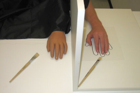

The rubber hand illusion (RHI) refers to the illusory perception of ownership of a rubber hand that may occur when covert tactile stimulation of a participant’s hand co-occurs with overt corresponding stimulation of a rubber hand. In the current study a team of IKN scientists from the Neuro- and Biopsychology labs investigated the influence of the RHI on pseudoneglect on the line bisection task (the leftward bias when marking the centre of horizontal lines). The results of study suggest that integrating the left sided rubber hand into one’s body image shifts the subjective body midline to the right, thus counteracting pseudoneglect.


Appropriate attention levels are pivotal for cognitive processes, and individual differences in attentional functioning are related to variations in the interplay of neurotransmitters. In the present study, the role of variation in GRIN2B, which encodes the NR2B subunit of N-methyl-d-aspartate (NMDA) receptors, was explored.
The study focuses on the regulation of arousal and attention by comparing the efficiency of the three attention networks alerting, orienting and conflicting as measured with the Attention Network Test (ANT). Two synonymous SNPs in GRIN2B, rs1806201 (T888T) and rs1806191 (H1178H) were genotyped in 324 young Caucasian adults. Results revealeda highly specific modulatory influence of SNP rs1806201 on alerting processes with subjects homozygous for the frequent C allele displaying higher alerting network scores as compared to the other two genotype groups (CT and TT). This result can be further explained by faster reaction times in the no-cue condition of the ANT (tonic alertness) in participants carrying at least one of the rare T alleles, possibly as a result of more effective glutamatergic neurotransmission.

Christian Beste successfully habilitated on the 10th of July 2012 in the Faculty of Psychology for “Cognitive Neuroscience”. His written thesis was already impressive by its coverage, depth, and publication quality. Now, he performed equally well with his oral presentation on “Effortless Learning: Fact or Fiction”. Christian could show that mere, high-frequency exposure learning can create an LTP-like process that dramatically increases the probability to detect certain stimuli. Conversely, an LTD-like training protocol decreases stimulus detection. Thus, effortless learning seems to work and thus is a fact. However, Christian also revealed that it is fiction to hope for a generalized effortless learning ability. Altogether, this was a very strong presentation and the committee unanimously decided that Christian Beste should be awarded with the habilitation of the Faculty of Psychology.
CONGRATULATIONS CHRISTIAN !!!


Dopamine D1-like receptors consist of D1 (D1A) and D5 (D1B) receptors and play a key role in working memory. However, their possibly differential contribution to working memory is unclear. In birds, the D1-like receptor family is extended and consists of the D1A, D1B, and D1D receptors. Their possibly differential contribution to cognitive training is mostly unclear. Biopsychologists from Bochum and Düsseldorf combined a working memory training protocol with a stepwise increase of cognitive subcomponents and real-time RT-PCR analysis of dopamine receptor expression in pigeons to identify molecular changes that accompany training of isolated cognitive subfunctions. They conducted a behavioral working memory paradigm that works like Russian Matryoshka dolls: The four tasks were designed with increasing cognitive demands such that task 2 had one cognitive component more than task 1, task 3 had more components than task 2 and so on. By subtraction of cognitive faculties between tasks, expression changes ofD1A, D1B, andD1D in striatum and avian "prefrontal cortex", could therefore be mapped to specific subcomponents of cognitive training. The data show that D1B receptor plasticity follows a training that includes active mental maintenance of information, whereas D1A and D1D receptor plasticity in addition accompanies learning of stimulus-response associations. Plasticity of D1-like receptors plays no role for processes like response selection and stimulus discrimination. None of the tasks altered D2 receptor expression. The present study shows for the first time that different cognitive components of working memory training have distinguishable effects on D1-like receptor expression.

Anna Ball successfully graduated on the 3rd of July 2012 and was awarded with a Dr. rer. nat in Psychology. Anna conducted a huge, highly complex and perfectly designed study in which she analyzed the alterations of hormones, brain asymmetries, psychophysical markers, social cognitive features, and motivational scores during the menstrual cycle. Anna departed in her study from an evolutionary perspective in which she proposed that cognitive changes during the menstrual cycle have to be seen as behavioral adaptations that increase the fitness of the individual. Going even a step further, she also assumed that alterations of brain asymmetries could represent the mean to achieve these behavioral changes. Anna tested three groups of subjects: normally cycling females, young female subjects that use a vaginal contraceptive ring (thereby preventing a normal cycle), and young men. In the end, she was rewarded with a large number of results that show how alterations of hormones affect various alterations of cognition, motivation, brain states, and strategic behavior. The committee was truly impressed with Anna's contributions and decided to award her the doctorate title with the grade magna cum laude.
CONGRATULATIONS ANNA !!!
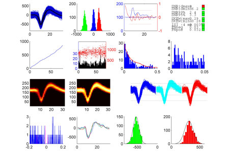

MLIB is a MATLAB-based software package for the analysis of the spike data, ie shapes and temporal patterns of extracellularly recorded action potentials. In particular, MLIB contains functions for a) assessing spike sorting quality / unit isolation, and b) constructing all sorts of peri-stimulus time histograms as well as raster displays and spike density functions constructed with various filter kernels. The software package is accompanied by an extensive documentation, along with many examples using spike data from operantly conditioned, freely moving pigeons. The software can be downloaded from Matlab Central File Exchange.
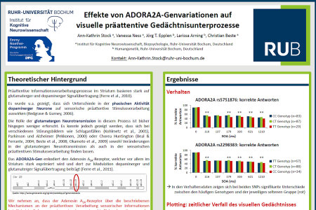

Ann-Kathrin Stock participated in this year’s “Psychologie und Gehirn” conference of the DGPA in Jena where she presented two posters. For one of her posters dealing with the differential effects of ADORA2A gene variations in pre-attentive visual sensory memory subprocesses she won the conference’s poster prize (“Posterpreis der Fachgruppe ‘Biologische Psychologie und Neuropsychologie’ der Deutschen Gesellschaft für Psychologie (DGPs) 2012”) endowed with a prize money of 300€.
The ADORA2A gene encodes the adenosine A2A receptor which is highly expressed in the striatum where it plays a role in modulating glutamatergic and dopaminergic transmission. While knowledge about the influence of phasic dopaminergic modulation on pre-attentive sensory stimulus processing is rather well-established, less is known about glutamatergic signaling in this context. Studying two polymorphisms, it could be shown that the rare homozygous genotypes seem to be associated with differences in the efficiency of pre-attentive sensory memory sub-processes most likely modulated by dopaminergic and glutamatergic signaling in a dissociable manner.
CONGRATULATIONS ANN-KATHRIN !!!
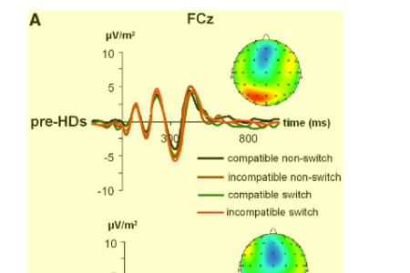

Flexible response adaptation and the control of conflicting information play a pivotal role in daily life. Yet, little is known about the neuronal mechanisms mediating parallel control of these processes. We examined these mechanisms using a multi-methodological approach that integrated data from event-related potentials (ERPs) with structural MRI data and source localisation using sLORETA. Moreover, we calculated evoked wavelet oscillations. We applied this multi-methodological approach in healthy subjects and patients in a prodromal phase of a major basal ganglia disorder (i.e., Huntington's disease), to directly focus on fronto-striatal networks. Behavioural data indicated, especially the parallel execution of conflict monitoring and flexible response adaptation was modulated across the examined cohorts. When both processes do not co-incide a high integrity of fronto-striatal loops seems to be dispensable. The neurophysiological data suggests that conflict monitoring (reflected by the N2 ERP) and working memory processes (reflected by the P3 ERP) differentially contribute to this pattern of results. Flexible response adaptation under the constraint of high conflict processing affected the N2 and P3 ERP, as well as their delta frequency band oscillations. Yet, modulatory effects were strongest for the N2 ERP and evoked wavelet oscillations in this time range. The N2 ERPs were localized in the anterior cingulate cortex (BA32, BA24). Modulations of the P3 ERP were localized in parietal areas (BA7). In addition, MRI-determined caudate head volume predicted modulations in conflict monitoring, but not working memory processes. The results show how parallel conflict monitoring and flexible adaptation of action is mediated via frontostriatal networks. While both, response monitoring and working memory processes seem to play a role, especially response selection processes and ACC–basal ganglia networks seem to be the driving force in mediating parallel conflict monitoring and flexible adaptation of actions.


It is widely held that spike responses of single neurons to sensory stimuli are noisy and uninformative. Accordingly, random fluctuations of individual neurons’ activity patterns are considered unimportant because downstream brain areas must perform some sort of averaging across large neuronal populations to obtain accurate information about sensory input variables. Contrary to this notion, recent studies have shown that electrical activation of single cortical neurons can indeed have behavioral relevance. For example, single-neuron stimulation in rat primary motor cortex generates movements of the facial whiskers. Moreover, stimulation of single neurons in somatosensory cortex yields a behavioral response in rats trained on a psychophysical detection task. These studies suggest that the influence of an individual cortical neuron on the local neural network is stronger than commonly thought. However, the mechanism by which the activity of a single neuron is amplified and propagated through the local neural network to eventually generate a behavioral response is unknown. The aim of the newly approved six-month research project is to begin to elucidate some of the mechanisms which underlie the phenomenon of single-cell induced behavioral responses. Together with Arthur Houweling at the University of Rotterdam, Maik will employ the juxtacellular stimulation technique to activate single neurons in the superficial layers of barrel cortex of anesthetized mice in a controlled manner while monitoring the activity of the local neural network.
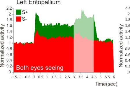

In humans and many other animals, the two cerebral hemispheres are partly specialized for different functions. However, knowledge about the neuronal basis of lateralization is mostly lacking. The visual system of birds is an excellent model in which to investigate hemispheric asymmetries as birds show a pronounced left hemispheric advantage in the discrimination of various visual objects. Therefore, biopsychologists and theoretical neuroscientists from Bochum and Freiburg, respectively, aimed to find a neuronal correlate for three hallmarks of visual lateralization in pigeons: first, the animals learn faster with the right eye–left hemisphere; second, they reach higher performance levels under this condition; third, visually guided behavior is mostly under left hemisphere control. To this end, Josine Verhaal (now at the Munich Technical University) recorded from the left and right forebrain entopallium while the animals performed a colour discrimination task. Josine found that, even before learning, left entopallial neurons were more responsive to visual stimulation. Subsequent discrimination acquisition recruited more neuronal responses in the left entopallium and these cells showed a higher degree of differentiation between the rewarded and the unrewarded stimulus. Thus, differential left–right responses are already present, albeit to a modest degree, before learning. As soon as some cues are associated with reward, however, this asymmetry increases substantially and the higher discrimination ratio of the left hemispheric tectofugal pathway would not only contribute to a higher performance of this hemisphere but could thereby also result in a left hemispheric dominance over downstream motor structures via reward-associated feedback systems.
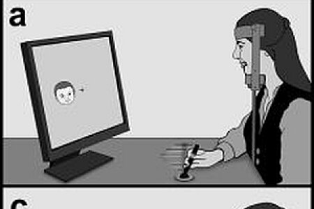

Possibly emotions are just a means to coordinate fast actions to stimuli that have a valence character. In the present study biological psychologists from Bochum and Würzburg planned to test if the left hemisphere preferentially controls flexion responses toward positive stimuli, while the right hemisphere is specialized toward extensor responses to negative pictures. To this end, right-handed subjects had to pull or push a joystick subsequent to seeing a positive or a negative stimulus in their left or right visual hemifield. Flexion responses were faster for positive stimuli (upper picture), while negative stimuli were associated with faster extensions responses (lower picture). Overall, performance was fastest when emotional stimuli were presented to the left visual hemifield. This right hemisphere superiority was especially clear for negative stimuli, while reaction times toward positive pictures showed no hemispheric difference. Thus, flexion and extension are indeed tightly linked to the valence of the stimulus. But the division of valence between hemispheres is not as clear. This fits to recent developments that an approach reaction (in this case visible as a flexion response) can be elicited by positive emotions as well as by anger, which is a negative emotion.


Smoking prevalence is highly elevated in schizophrenia compared to the general population and to other psychiatric populations. Evidence suggests that smoking may lead to improvements of schizophrenia-associated attention deficits; however, large-scale studies on this important issue are scarce. A group of psychiatrists from Berlin and biopsychologists from Bochum examined whether sustained, selective, and executive attention processes are differentially modulated by long-term nicotine consumption in 104 schizophrenia patients and 104 carefully matched healthy controls. Smoking was significantly associated with a detrimental conflict effect in controls, while the opposite effect was revealed for schizophrenia patients. Likewise, a positive correlation between a cumulative measure of nicotine consumption and conflict effect in controls and a negative correlation in patients were found. These results provide evidence for specific directional effects of smoking on conflict processing that critically dissociate with diagnosis. The data supports the self-medication hypothesis of smoking in schizophrenia and suggests selective attention as a specific cognitive domain positively targeted by nicotine consumption. The authors offer a model to explain the dissociating effects by nicotine-dopamine interactions located in the ventral tegmental area that lead to an increased prefrontal D1 receptor activation. As a consequence, smoking is disadvantageous for healthy participants with a priori favorable dopamine levels, but reinstates an advantageous D1 state in schizophrenia patients who otherwise suffer from a marked prefrontal dopamine deficit.
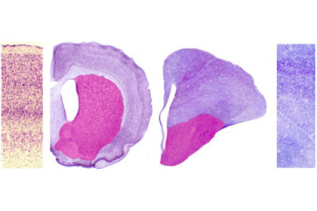

This review by Onur Güntürkün describes a case of convergence in the evolution of brain and cognition. Both mammals and birds can organize their behavior flexibly over time and evolved similar cognitive skills. However, mammals and birds display vast differences in the organization of their forebrains with mammals having a laminated cortex. The avian forebrain displays no such lamination; hence, lamination does not seem to be a requirement for higher cognitive functions. In mammals, executive functions are associated with the prefrontal cortex. The corresponding structure in birds is the nidopallium caudolaterale. Anatomic, neurochemical, electrophysiologic and behavioral studies show these structures to be highly similar, but not homologous. Thus, despite the presence (mammals) or the absence (birds) of a laminated forebrain, 'prefrontal' areas in mammals and birds converged over evolutionary time into a highly similar neural architecture. The neuroarchitectonic degrees of freedom to create different neural architectures that generate identical prefrontal functions seem to be very limited.
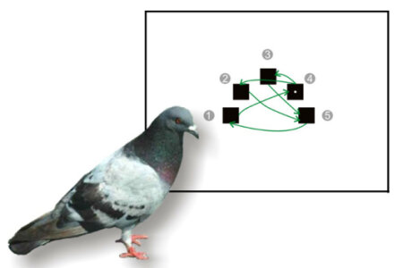

Most of our behavior is composed of action sequences, like driving a car, brushing your teeth or human language. The pigeon is a commonly used model organism to investigate the principles of how sequences are learned and executed. However, so far virtually nothing was known about the underlying neural circuits in the pigeon brain.
In a recent study scientists of the IKN identified two structures that play a role for the execution of a learned sequence in the pigeon.
The scientist applied a so called serial reaction time task (SRTT). This kind of task requires subjects to respond quickly to a stimulus that appears at five different locations in a fixed sequence. In their study the scientists transiently inactivated different brain areas of the pigeons during the execution of this sequential task. Results show that two regions play a central role. On the one hand, the nidopallium caudolaterale (NCL), that is considered the avian equivalent to mammalian prefrontal cortex. On the other hand, the nidopallium intermedium medialis pars laterale (NIMl). This area is comparable in some characteristics to a nucleus of the neural system that generates song in song birds. Hence, this study is the first to identify components of a neural system for the execution of sequential behavior in the pigeon.
Link: http://www.sciencedirect.com/science/article/pii/S0166432812001118
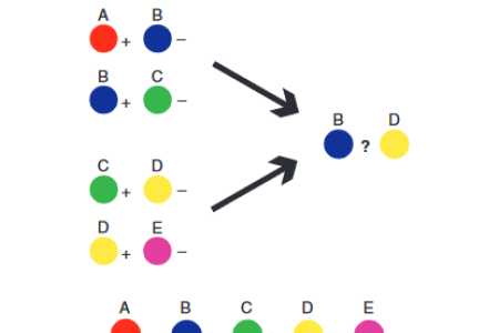

Specialization of the brain, where the left and right hemispheres perform different functions, is thought to have evolutionary advantages. But for many tasks, the hemispheres have to combine their expertise for optimal information processing.Scientists from the IKN studied hemispheric cooperation in pigeons, which have a visual system that develops asymmetrically in response to light stimulation. This allows comparison of integration capacities of lateralized (light-incubated) and non-lateralized (dark-incubated) animals. By presenting the left or right eye of the birds with different colour pairings, the scientists show that lateralized pigeons are able to integrate information learnt separately by the two hemispheres to solve a cognitive puzzle. In contrast, non-lateralized birds could not discriminate between colour pairings that required the combination of hemispheric-specific knowledge. This study provides first direct evidence that the efficiency of cooperation between the two brain halves depends on the development of hemispheric specialization, based on the experiences of the embryo.
Link: http://www.nature.com/ncomms/journal/v3/n2/full/ncomms1699.html#/abstract
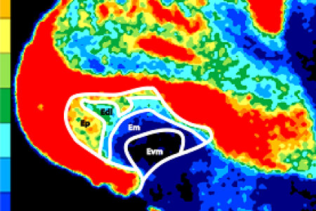

The 5-HT1A receptor is the most widespread serotonin receptor type and is involved in various functions like depression, schizophrenia, Alzheimer’s disease, and eating disorders. Our knowledge about the distribution of 5-HT1A receptors in birds is extremely limited. Therefore, a team of neuroanatomists and biopsychologists from the universities of Düsseldorf and Bochum analyzed the distribution of 5-HT1A receptor binding sites in the pigeon brain using quantitative in vitro receptor autoradiography with the selective radioligand [3H]-8-Hydroxy-2-(di-n-propylamino)tetralin ([3H]-8-OH-DPAT). What they discovered is a completely new landscape of neural subdivisions that hints to functional specializations that were unknown before. The picture of the entopallium shown here is a nice example for this new landscape that testifies the existence of new subdivisions. In addition, the regional pattern of distribution of 5-HT1A receptors displays a scattering similar to brain structures of mammals, furthering the discussion on the comparison of the avian and the mammalian brain.
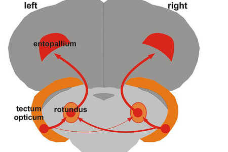

Hemispheric asymmetries play an important role in almost all cognitive functions. For more than a century, they were considered to be uniquely human but now an increasing number of findings in all vertebrate classes make it likely that we inherited our asymmetries from common ancestors. In a new review article in Frontiers in Comparative Psychology, IKN researchers explain how studying animal models could provide unique insights into the mechanisms of lateralization. Three avenues of research are outlined by providing an overview of experiments on left-right differences in the connectivity of sensory systems, the embryonic determinants of brain asymmetries, and the genetics of lateralization. All these lines of studies could provide a wealth of insights into human asymmetries that should and will be exploited by future analyses.
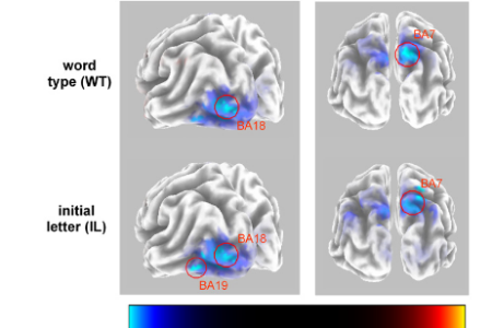

In the present study a team of IKN scientists investigated the relevance of hemispheric asymmetries for cognitive control processes using a lateralized version of the task switching paradigm.
ERPs were recorded and the neural sources of the ERPs were reconstructed using standardised low resolution brain electromagnetic tomography (sLORETA).
A left lateralization of the N1 that was mediated by activation in the left extrastriate cortex as well as a greater positivity of the P3b were observed after stimulus presentation in the
RVF compared to the LVF. These findings reveal that FCAs are an important modulator of executive functions related to cognitive flexibility.


There is growing interest in understanding the neurobiological foundations of attention.
Since dopamine is one of the most important neurotransmitters regulating attention processes, we aimed to examine whether attentional processes in a change detection task (biased competition paradigm) are modulated by dopamine signaling. Therefore, we investigated the influence of two polymorphisms of the dopaminergic system, i.e., Val158Met (rs4680) in the catechol-O-methyltransferase (COMT) and a variable number of tandem repeats polymorphism (VNTR, rs28363170) in the dopamine transporter (DAT1). Based on previous research, it is known, that the COMT Met allele results in lower enzyme activity and is therefore related to enhanced PFC dopamine signalling. In this study with 216 Caucasian subjects, we found that homozygous Met/Met allele carriers had difficulties when performing the biased competition task, particularly showing the greatest difficulties in case cognitive and behavioural flexibility was necessary and the required reaction was not part of the subject’s primary task set.
Contrary, no differences between the two genotype groups were evident, when an attentional conflict emerged and attentional control was needed for adequate responding.
With respect to other studies examining mechanisms of attentional functions in different paradigms, the results suggest that behavioural flexibility and attentional control as two execeutive subprocesses are differentially influenced by genetic polymorphisms within the dopaminergic system.

From 4th to 23rd of September 2011 we had the great pleasure of hosting the first “European Campus of Excellence” in Neuroscience at the Ruhr University Bochum. Over a period of three weeks thirty truly gifted students from all over Europe visited Bochum to deepen their knowledge in memory research. The present volume is a compilation of many of those ECE memories proving that our research is not only relevant to understanding human behavior but can also be a lot of fun for students, scholars and laymen. Enjoy Reading!


Our seamless stream of thoughts and actions cloud be one of the greatest mysteries of the mind. This mystery is the ease with which we constantly and effortlessly chose one thing to do and many other things to let. Even more miraculously, we sometimes switch to a mode in which we can work in parallel on some tasks to then switch back to a serial mode of working. Often this flexibility of our mind was discussed in a corticocentric way, as if our cortex does it all. Now, the newly accepted and DFG-funded Emmy Noether group of Christian Beste set sails to completely change perspectives. In a grand effort that involves psychological, genetic, neurological, electrophysiological, theoretical, and imaging-based approaches, he and his team is dedicated to find crucial answers to the mysteries of the parallel and/or serially organized mind by focusing on the intricacies of the basal ganglia. The reviewers of the DFG were deeply impressed with the 5 year plan and decided to generously support this endeavor. By acquiring an Emmy Noether group, Christian is eligible to create his own independent research unit that is associated with the Biopsychology lab.
CONGRATULATIONS CHRISTIAN !!!


The overwhelming majority of research in the neurosciences employs p-values stemming from tests of statistical significance to decide on the presence or absence of an effect of some treatment variable. Although a continuous variable, the p-value is commonly used to reach a dichotomous decision about the presence of an effect around an arbitrary criterion of 0.05. This analysis strategy is widely used, but has been heavily criticized in the past decades. To counter frequent misinterpretations of p-values, methodologists advocate complementing or replacing p-values with measures of effect size (MES). Many psychological, biological, and medical journals now recommend reporting appropriate MES. One hindrance to the more frequent use of MES may be their scarcity in standard statistical software packages. Also, the arguably most widespread data analysis software in neuroscience, MATLAB, does not provide MES beyond correlation and receiver-operating characteristic analysis. In our article, we review the most common criticisms of significance testing and provide several neuroscience examples where usage of MES conveys insights not amenable through the use of p-values alone. We introduce an open-access MATLAB toolbox providing a wide range of MES to complement the frequently used types of hypothesis tests, such as t-tests and analysis of variance. The accompanying documentation provides calculation formulae, intuitions for interpretation, and example calculations for each measure. The toolbox described in this article is usable without sophisticated statistical knowledge and should be useful to neuroscientists wishing to enhance their repertoire of statistical reporting.

Conny Hahn successfully graduated on the 28th of November 2011 at the IGSN and was awarded with a PhD in Neuroscience. Conny submitted a thesis consisting of four papers on the effects of smoking on language asymmetry and attention. She could show that smoking, possibly via the cellular effects of nicotine, is able to alter functional brain asymmetries and attentional resources. Some of these effects show a clear sex-dependency. Most interestingly, Conny was able to show that if one controls for cigarette consumption, asymmetries in schizophrenic patients are no different from controls. Thus, some of the effects of schizophrenia on cerebral asymmetries might arise due to the heavy nicotine consumption of these patients. The international committee was impressed with Conny's contributions and decided to award her the title PhD of Neuroscience with the grade magna cum laude.
CONGRATULATIONS CONNY !!!

Sebastian Ocklenburg successfully graduated on the 25th of November 2011 in the Faculty of Psychology. He so much impressed the committee with his outstanding thesis and his strong defense that they unanimously decided to grade his doctoral project with a summa cum laude. Given the high threshold for this grade in our Faculty, this is an extremely rare and very prestigious event. Sebastian submitted a publication-based thesis with six papers in which he analyzed the phylogenetic, ontogenetic, genetic, and neuronal mechanisms of cerebral asymmetries. He was able to show that vocal asymmetry has a long evolutionary history, that experience-based factors shape human handedness, and that early neuronal processes guided language lateralization. In addition, he identified a genetic association for language asymmetry.
CONGRATULATIONS SEBASTIAN !!!


Callosal agenesis is a condition in which the Corpus callosum – the largest nerve fiber bundle connecting the two cerebral hemispheres – is developmentally absent due to genetic or environmental factors. Although this implies an absence of more than 180 million nerve fibers, past research revealed that the brain’s reorganization power may allow for a compensation for this malformation in certain unisensory tasks, e.g. by an increased use of other pathways such as the anterior commissure. Until now, however, potentials and limits of the brain’s plasticity have not been investigated in experimental setups which involve interactions between different sensory modalities. Therefore, a team of Biopsychologists from Hamburg and Bochum investigated five individuals with callosal agenesis in a visuotactile interaction task, the Crossmodal Congruency Task, which involves speeded tactile judgments in accompaniment of visual distractors. Results showed that interactions between vision and touch did not differ between individuals with callosal agenesis and healthy controls. This implies that the Corpus callosum is not necessary for visuotactile perception per se, and that early reorganization mechanisms may compensate for the absence of the corpus callosum.
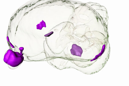

There is something mysterious about olfaction. It is the only sense that directly enters the forebrain without a thalamic relay. It also has tight links with the limbic system; creating the myth that olfaction is a strong gateway into emotional memory. Pigeons also use olfactory cues to navigate over unfamiliar areas, and any impairment of the olfactory system generates remarkable reduction of homing performance. This special function of olfaction is also lateralized: Studies suggest a critical involvement of the right olfactory bulb (OB) and the left piriform cortex (CPi) for initial orientation. Unfortunately, the structural organization of the olfactory system is by far not clarified yet. Thus, a team of Biopsychologists from Bochum and Witwatersrand (South Africa) re-analyzed the system by antero- and retrograde tract tracing with biotinylated dextran amine and choleratoxin subunit B, and especially evaluated quantitative differences in the number of cells in the OB innervating the left and right CPi. They verified a strong bilateral input to the CPi, and the prepiriform cortex (CPP), as well as small projections to the ipsilateral medial septum and the dorsolateral corticoid area and the nucleus taeniae of the amygdala in both hemispheres. The adjacent picture depicts some of the olfactory components within a 3D-view of the pigeon brain. However, the authors could not reveal any asymmetries within in the projections described. Thus, the functional lateralization of the olfactory system is not simply based on differences in the number of projecting axons of the major processing streams.
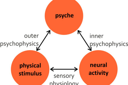

Single-unit recordings conducted during perceptual decision-making tasks have yielded tremendous insights into the neural coding of sensory stimuli. In such experiments, detection or discrimination behavior (the psychometric data) is observed in parallel with spike trains in sensory neurons (the neurometric data). Frequently, candidate neural codes for information read-out are pitted against each other by transforming the neurometric data in some way and asking which code’s performance most closely approximates the psychometric performance. The code that matches the psychometric performance best is retained as a viable candidate and the others are rejected. In following this strategy, psychometric data is often considered to provide an unbiased measure of perceptual sensitivity. It is rarely acknowledged that psychometric data result from a complex interplay of sensory and non-sensory processes and that neglect of these processes may result in misestimating psychophysical sensitivity. This again may lead to erroneous conclusions regarding the adequacy of neural candidate codes. In this review, we first discuss requirements on the neural data for a subsequent neurometric-psychometric comparison. We then focus on different psychophysical tasks for the assessment of detection and discrimination performance and the cognitive processes that may underlie their execution. We discuss further factors that may compromise psychometric performance and how they can be detected or avoided. We believe that these considerations point to shortcomings in our understanding of the processes underlying perceptual decisions, and therefore offer potential for future research.
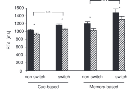

In this study we examined the relevance of the functional brain-derived neurotrophic factor (BDNF) Val66Met polymorphism as a modulator of task-switching performance in healthy elderly by using behavioral and event-related potential (ERP) measures. Task switching was examined in a cue-based and a memory-based paradigm. Val/Val carriers were generally slower, showed enhanced reaction time variability and higher error rates, particularly during memory-based task switching than the Met-allele individuals. On a neurophysiological level these dissociative effects were reflected by variations in the N2 and P3 ERP components. The task switch-related N2 was increased while the P3 was decreased in Met-allele carriers, while the Val/Val genotype group revealed the opposite pattern of results. In cue-based task-switching no behavioral and ERP differences were seen between the genotypes. These data suggest that superior memory-based task-switching performance in elderly Met-allele carriers may emerge due to more efficient response selection processes. The results implicate that under special circumstances the Met-allele renders cognitive processes more efficient than the Val/Val genotype in healthy elderly, corroborating recent findings in young subjects.


Fronto-striatal loops play an important role action selection processes, especially when discordant sensory and contextual information has to be integrated to allow adequate selection of actions. Yet, it is widely unknown how far changes in the precision of neural synchronization processes are induced by only slight dysfunctions of striatal neural inter-connectivity and in how far such slight changes may affect action selection processes. To elucidate the role of fronto-striatal interactions, these processes may be examined in neurodegenerative basal ganglia disorders. We investigated these processes in a neurogenetic, degenerative disorder (i.e. Huntington's disease) in a modified Go/Nogo task, while assessing neural synchronization processes by means of phase-locking factors (PLFs) as derived from event-related potentials (ERPs). The results show that gene mutation carriers, who have not yet developed clinical signs of this movement disorder (i.e., pre-HDs) only encounter problems in response inhibition, when discordant contextual information and sensory input have to be integrated. No deficits were evident, when response inhibition can be based on more habitual stimulus–response mappings, i.e., when contextual and sensory information were congruent. While 'habitual' action selection is unaffected by changes in striatal structures influencing reliability of neural synchronization processes, efficient 'controlled' processes of action seem to be closely dependent upon highly reliable neural synchronization processes. The neurophysiological analysis suggests that especially pre-motor inhibition processes (Nogo-N2) are affected. This was most strongly reflected in a decline in the degree of phase-locking in the Nogo-N2 range. Deficits in pre-HDs seem to emerge as a consequence of phase-locking-behavioural decoupling. Of clinical interest, declines in the precision of phase-locking depended on the amount of the individual's mutant huntingtin exposure and predicted the probability of disease manifestation in the next five years. This suggests that phase-locking parameters may prove useful in future studies evaluating a possible function as a biomarker in Huntington's disease.


In perceptual decision making tasks, subjects are required to sort a range of stimuli into two or more categories. From the viewpoint of the experimenter, this is a psychophysical task: the subject tries to decide on the basis of sensory evidence which category a given stimulus belongs to. Thus, the subject is assumed to attempt to maximize accuracy. However, when the subjects are animals, conditions may change: to perform well in a behavioral experiment, animals are usually rewarded for each correct response. Thus, from the viewpoint of the animal subject, the task is more about foraging than about psychophysics.
In a newly released paper, Maik Stüttgen, Ali Yildiz and Onur Güntürkün report that rewarding animal subjects for one kind of classification more often than for another kind heavily biases their choice behavior. More importantly, the animals were shown to integrate sensory evidence - which category the stimulus belongs to - and reward preference - which category response is rewarded more often - in a statistically optimal fashion. Moreover, the subjects did so in a suprisingly brief amount of time, which calls into question that they attempted to maximize expected value as decision goal - a central assumption of signal detection theory.
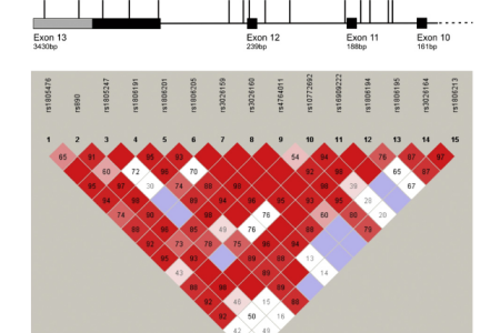

In recent studies by our group we accumulated evidence that the effect of certain genetic polymorphisms varies across cognitive processes. The polymorphisms 5-HT1A C(-1019)G (Beste et al., 2011) and TNF-alpha -308G-A (Beste et al., 2011) have been shown to participate in the modulation of cognitive control. For the first time, the present study provides evidence that variations in the GRIN2B gene are associated with decision making.The dopaminergic system is known to modulate decision-making. As N-methyl-d-aspartate (NMDA) receptors strongly influence dopaminergic function, it is conceivable that the glutamatergic system is also involved in decision-making. We examined whether polymorphisms in the N-methyl-d-aspartate receptor 2B subunit gene (GRIN2B) influence decision-making using the Iowa Gambling Task (IGT). In total, 245 (n = 245, 127 female) healthy German students were included in the analysis. Two synonymous SNPs in exon 13, rs1806191 (H1178H) and rs1806201 (T888T) showed the strongest association with aspects of IGT performance. Females with a CC allele in rs1806201 made less use both of a win-stay strategy and demonstrated more exploratory behaviour during task execution. For rs1806191, we found a strong additive effect in usage of a win-stay strategy. This, partly sex-dependent, correlation of the win-stay/lose-shift behaviour with GRIN2B genotypes suggests that healthy individuals with certain GRIN2B variations respond differently to ambiguous conditions, possibly by altered perception of wins and losses. These findings underline the necessity to integrate the glutamatergic system when examining decision-making processes.
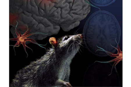

In the current study, a team of scientists from the IKN and the Department of Human Genetics investigated whether variations in the N-methyl-d-aspartate receptor 2B subunit gene (GRIN2B) influence language lateralization and handedness in healthy individuals. In a cohort of 424 genetically unrelated participants a significant association between the synonymous GRIN2B variation rs1806201 and language lateralization assessed using the dichotic listening task, but not handedness, was observed. These findings suggest for the first time that variation in NMDA-receptors contributes to the interindividual variability of language lateralization.
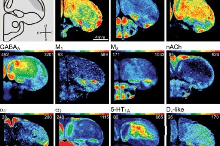

Mapping a territory is always the first step of analysis. So, mapping the nidopallium caudolaterale (NCL), an avian functional analogue to the mammalian prefrontal cortex, in 1993 was a prerequisite for subsequent research. However, a more comprehensive and truly comparative mapping was meanwhile needed. Therefore, scientist from the Institute of Brain Research in Düsseldorf and Biopsychologists from Bochum analyzed binding site densities of AMPA, NMDA, Kainite, GABAA, M1, M2, and nicotinic nACh, a1, a2, 5-HT1A, and D1-like receptors using quantitative in vitro receptor autoradiography. They compared the receptor architecture of the pigeons’ NCL, with prefrontal areas in rats and humans. Their findings enable a novel delineation of the avian NCL from surrounding structures and a further parcellation into medial and lateral components. Comparisons of the NCL with the rat and human frontal structures showed differences in the receptor distribution, particularly for glutamate receptors, but also revealed highly conserved features like for GABAA, M2, nACh and D1-like receptors. Assuming a convergent evolution of avian and mammalian prefrontal areas, these results support the hypothesis that specific neurochemical traits provide the molecular background for higher order processes such as executive functions.


The board of trustees of the Attempto Foundation of the University of Tübingen has decided to award Maik Stüttgen this year's Attempto Award for his recent paper, "Integration of vibrotactile signals for whisker-related perception in rats is governed by short time constants: comparison of neurometric and psychometric performance", appeared in The Journal of Neuroscience last year.
The paper investigated mechanisms of sensory coding in the primary somatosensory ("barrel") cortex of the rat. The animals' ability for detecting multi-pulse tactile stimuli was measured simultaneously with the activity of single neurons in barrel cortex. The authors constructed a theoretical model which successfully relates neural response patterns to both detection probabilities and reaction times of the animals, thus bridging the gap between neural activity on the one hand and sensation and perception on the other.
The award ceremony will take place on October 12th Tübingen.


In recent studies by our group we accumulated evidence that the effect of certain genetic polymorphisms varies across cognitive processes. The results call in question, if a ‘risk allele’ necessarily confers a disadvantage to its carriers. In this study we provide further evidence on this issue.
Cognitive control processes may depend on contextual information, sometimes improving performance, but impairing performance if expectancies about forthcoming events induce pre-potent responses. The neurobiological bases of these effects are not understood. Here, we examine context-dependent variations of response control processes using the AX-CPT task with respect to the relevance of the functional serotonin 1A receptor polymorphism (5-HT1A C(−1019)G). The results show that, when context information is helpful to drive behavioural performance, carriers of the −1019G allele reveal compromised cognitive control. Yet, they show enhanced task performance when strong context representations would lead to declines in behavioural control. These findings are paralleled by modulations of the (Nogo)-P3 ERP-component. These results show for the first time that, even though the −1019G allele enhances the risk to develop anxiety disorders, it also confers an advantage to its carriers in terms of better cognitive control processes in conditions where contextual information compromises cognitive control. Effects of the 5-HT1A C(−1019)G polymorphism were further modulated by anxiety sensitivity. As the functional effect of the 5-HT1A C(−1019)G polymorphism has previously been shown to be rather specific for serotonergic 1A autoreceptors in the dorsal raphe nucleus (DRN), the results suggest that contextual modulations in cognitive control may be exerted by the DRN.


Was ist eigentlich das Gehirn und wie funktioniert es genau? Am 10. Juli bietet das IKN, im Rahmen des von der "Sendung mit der Maus" organisiertem Türöffner Tag, einen Workshop an, in dem Kinder mehr über das wohl wichtigste Organ unseres Körpers erfahren können. Zusammen mit Forschern und Studenten des IKN werden die Besucher Gehirnmodelle aus Knete anfertigen. Natürlich können alle Fragen zum Thema "Gehirn und Nervensystem" gestellt werden.
Das Institut öffnet seine Tür am 10. Juli ab 14 Uhr für Kinder ab 4 Jahren.
Ort: Ruhr-Uni-Bochum, Universitätsstraße 150, Institut für kognitive Neurowissenschaften, GAFO 05/609. Die Veranstaltung dauert 1 bis 2 Std. Anmeldungen bitte per Mail an: felix.stroeckens@rub.de

Maik Stüttgen has been granted a research project on the neural basis of adaptive decision-making. The project will be funded for three years and will focus on neural correlates of subjective value.
Classical models of decision making assume that the objective value of different choice options is transformed to subjective value, i.e. the expected utility of a choice outcome as assessed by an individual subject. Subjective value is known to be influenced by a variety of factors, such as the magnitude of a gain or a loss. Recent studies have suggested that subjective value is coded by the spiking activity of individual neurons in forebrain areas. However, little is known about the properties of the value representation when negative consequences, such as loss of reinforcers, are encountered. Furthermore, the plasticity of these representations has not been characterized. In the present project, pigeons will be exposed to complex perceptual discrimination tasks in a non-stationary environment. Concomitant unit recordings in forebrain areas will enable to relate adaptive decision making to changes in neuronal coding properties.

Cellular studies have focused on long-term potentiation (LTP) and long-term depression (LTD) to understand requirements for persistent changes in synaptic connections. Here we adapted LTP/LTD-like protocols to visual stimulation to alter human visual attention and behavior. In a change-detection task, participants reported luminance changes against distracting orientation changes. Subsequently, they were exposed to passive visual high- or low-frequency stimulation of either the relevant luminance or irrelevant orientation feature. LTP-like high-frequency protocols using luminance improved ability to detect luminance changes, whereas low-frequency LTD-like stimulation impaired performance. In contrast, LTP-like exposure of the irrelevant orientation feature impaired performance, whereas LTD-like orientation stimulation improved it. This is the first study showing that mechanisms used in cellular studies can also be used in vivo to alter human behavior and supports cellular, neurophysiological studies showing that these mechanisms alter synaptic strength. Future research may evaluate the potential of this technique for clinical interventions.
Comment on this article:


Onur Güntürkün becomes a member of the Editorial Board of Behavioural Processes (Elsevier). Behavioural Processes publishes research on animal behavior from behavioral analytic, cognitive, ethological, ecological and evolutionary points of view. The journal plans to make a strategic shift to incorporate contributions from neuroscience without losing its strength in the field of ethology-minded behavioral analyses. Troubled waters; Onur Güntürkün was seen as a person to help in this transition.

Schizophrenia has been associated with deficits in functional brain lateralization and some authors even argue that the reduction of asymmetry produces the psychosis. However, there is one major and often overlooked confound: Schizophrenic patients are extreme cigarette smokers. This association is very interesting, because a team from the Bochum Biopsychology Department could recently show that smoking can reduce auditory language asymmetry. Thus, the altered laterality pattern in schizophrenia could, at least in part, result from secondary artifacts due to smoking rather than being a pure cause of the disease itself. To test this hypothesis, the present study examined auditory language lateralization in 67 schizophrenia patients and in 72 healthy controls. Again it was found that smoking reduces language lateralization. Most importantly, no further effect of schizophrenia on language asymmetry was found. This opens the tantalizing possibility that at least some of the asymmetry alterations in schizophrenia are not a primary consequence of the disease but a secondary effect of increased smoking habits.
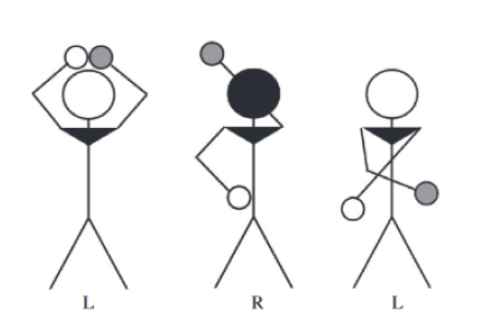

Several studies have demonstrated that women believe they are more prone to left–right confusion than men.However, while some studies report that there is also a sex difference in LRC tasks favouring men, others report that men and women perform equally well.Recently, it was suggested that sex differences only emerge in LRC tasks when they involve mental rotation.To test this assumption, a team of biopsychologists from Durham University (Durham, England) together with biopsychologists from Bochum tested 91 participants with two LRC tasks.To rule out the possibility that sex differences in LRC are confounded by sex differences in mental rotation, male and female participants were matched for mentalrotation performance. These matched participants showed robust sex differences in favour of men in all LRC measurements.


On the 7th of April 2011, Onur Güntürkün was awarded in Düsseldorf with the Medal of Merit of North Rhine-Westphalia. As explained by Minister-President Hannelore Kraft in her laudatio, the medal was awarded for his scientific success and his contribution to the academic exchange between Germany and Turkey.
With april the 22nd Christian Beste has been appointed as a member of the Editorial Advisory Board of Neuropsychologia. Neuropsychologia is a prestigeous, traditional journal in the field of cognitive neuroscience. Together with the other members of the Editoral Advisory Board his task is to further improve journal standards by monitoring the editorial policy of the journal in terms of coverage, level and quality of the papers.
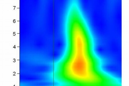

In the present study a team of IKN scientists investigated the temporal and spectral dynamics as well as the cortical networks underlying the interaction of hemispheric asymmetries and executive functions related to response inhibition.To this end, event-related potentials were recorded during tachistoscopic presentation of verbal 'Go' and 'Nogo' stimuli in the left (LVF) and theright visual field (RVF). Participants committed fewer false alarms to verbal Nogo stimuli presented in the RVF than to stimulipresented in the LVF. This asymmetry was paralleled by the neurophysiological data. The Nogo-N2 and relateddelta frequency band power were stronger when response inhibition was driven by stimuli presented in theLVF, implying a stronger response conflict. This effect was mediated by stronger activations in bilateralmedial-prefrontal and especially left parietal networks. These findings show that hemispheric dominances in information processingcan induce differences in demands on cognitive processes operating via bilateral networks that ultimatelydrive behavioural asymmetries.


From September 4th – 25th, 2011, the first ECE course in neuroscience will be held at the Ruhr-University Bochum. The ECE is promoting a European network of summer schools that bring eminent scientists and 30 highly talented and gifted European (plus Israel, Norway, Switzerland, and Turkey) students together. Central theme of the summer course in Bochum is: "The Fate of the Memory Trace: Learning, remembering and forgetting from molecules to behavior." Over a period of 3 weeks the students attend lectures in the morning and practical lab excercises in the afternoon.The topics of the first week are "Acquisition and Consolidation". In week 2 the students deepen their knowledge on "Retrieval and the Memory System". Week 3 is dedicated to "Extinction and Forgetting". At weekends our guests get to know more about the Ruhr area and, of course, each other. The Campus in Bochum is possible due to the generous funding of Stiftung Mercator. The selected students receive stipends covering travel, housing and full board. Onur Güntürkün is in charge of organizing the summer school.


In patient studies, impairments of sense of body ownership have repeatedly been linked to right-hemispheric brain damage.In the current study a team of IKN scientists from the Neuro- and Biopsychology labs investigated lateralisation for sense of body ownership in healthy adults.To this end the rubber hand illusion was elicited on both hands of left- and right-handed participants.The vividness of the illusion was measured by subjective self-reports as well as by skin conductance responses to watching the rubber hand being harmed.Handedness did not affect the vividness of the illusion, but a stronger skin conductance response was observed, when the illusion was elicited on the left hand.These findings suggest a right-hemispheric dominance for sense of body ownership in healthy adults.
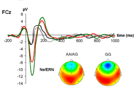

Neuroimmunological factors may modulate brain functions and are important to understand the molecular basis of cognition. Previously, we have been shown that these factors modulate functions mediated by different cortical areas in a dissociated fashion. In the current study we ask, if such dissociated effects are also evident between adjacent functional fronto-striatal loops. Since the basal ganglia are neurobiologically heterogeneous, different cognitive functions mediated by basal ganglia-prefrontal loops (response inhibition and error processing) may not necessarily be uniformly affected. Response inhibition and error processing functions were examined using event-related potentials (ERPs) and subjects were genotyped for the functional TNF-alpha -308G-A polymorphism. We show a double-dissociated effect of the functional TNF-alpha -308G-A polymorphism on response inhibition and error processing. While response inhibition functions were more effective in the AA/AG genotype group, error monitoring functions are adversely affected in this genotype group. In the GG genotype group, the pattern of results was vice versa. The results refine the view of the effects of TNF-alpha on cognitive functions.
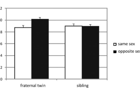

Men outperform women in mental rotation by about one standard deviation. Prenatal exposure to testosterone has been suggested as one cause. In animals it has been shown that a female fetus located between two male ones is exposed to higher levels of testosterone. It is still unclear whether intra-uterine hormone transfer exists in humans. Therefore, the influence of an intra-uterine presence of a male co twin was studied in female fraternal twins (N=200). Women with a male co-twin outperformed women with a female co-twin by about a third standard deviation. In a no-twin control group (N=200), performance of women with a slightly older sibling did not depend upon the sibling's sex. These findings provide preliminary support for the theory of an influence of prenatal testosterone on mental rotation performance.


The brain-derived neurotrophic factor (BDNF), a member of the neurotrophin family, is involved in nerve growth and survival. Especially, a single nucleotide polymorphism (SNP) in the BDNF gene, Val66Met, has gained a lot of attention, because of its effect on activity-dependent BDNF secretion and its link to several cognitive functions, especially memory processes. In this study we investigated the functional relevance of BDNF for pre-attentive visual sensory memory processes. Since BDNF is also discussed to be involved in the pathogenesis of depression, we additionally tested for possible interactions with depressive mood. The BDNF Val66Met polymorphism significantly influences iconic-memory performance, with the combined Val/Met-Met/Met genotype group revealing less time stability of information stored in iconic memory than the Val/Val group. Furthermore, this stability was positively correlated with depressive mood exclusively in the Val/Val genotype group. Thus, these results show that the BDNF Val66Met polymorphism has an effect on pre-attentive visual sensory memory processes and also suggests that sensory memory processes may proof useful in the search for cognitive endophenotypes of depression, which may be target for future studies.
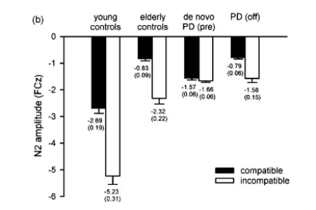

Action selection mechanisms depend upon fronto-striatal structures and are impaired in neurodegenerative basal ganglia disorders. Dopaminergic treatment of Parkinson's disease lingers clinical symptoms, but the question what effects these treatments have neurophysiological mechanisms underlying complex action selection processes have only rarely been addressed.We examined effects of short-term and long-term dopaminergic medication in Parkinson's disease on conflict monitoring or response selection processes. These processes were examined using event-related potentials (ERPs), while subjects performed a stimulus-response (S-R) compatibility task. An extended sample of young and elderly controls, Parkinson's disease patients with a medication history (PDs) and initially diagnosed, drug-naïve de novo PD patients (de novo PDs) were enrolled. Both PD groups were measured twice (on and off-medication or before and 8 weeks after medication onset). The results show that dopaminergic intervention selectively reduced the pathologically enhanced response selection in compatible S-R relations. This medication effect was already evident after short-term treatment, not differing from long-term treatment and performance in elderly controls. Contrary, age-related attenuations of the N2 in incompatible S-R relations, probably reflecting impaired conflict processing or response control, are unaffected by medication. The results suggest that compatible and incompatible S-R relations demand different neuronal mechanisms within the basal ganglia, as only the former are affected by agonizing the dopaminergic system.

We can learn new information and are subsequently able to remember it. However, we are equally able to learn that once acquired information is no longer valid and cease to respond to it. While the initial process of acquisition of new knowledge is well studied, the process of extinction is far less understood. It is known that extinction involves a new learning process that is different and more complex than the initial acquisition of the CS-US-association. Extinguished responses do not disappear but can return following manipulations such as a change in context as in renewal paradigms. Within the scope of the Research Unit that is going to start working on 1st of January 2011, 8 labs from the universities of Bochum, Essen-Duisburg and Marburg plan to explore the neural, the behavioral, and the clinical mechanisms of extinction in various species, incl. humans. Using a highly interactive research strategy, they intend to harvest deep insights into both the common and the distinct mechanisms of extinction learning in different systems and organisms. By this, there is good hope to achieve translational insights between Basic and Clinical Science. Onur Güntürkün is the speaker of the Research Unit.
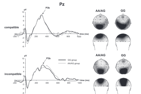

There is growing interest to understand the molecular basis of complex cognitive processes. While neurotransmitter systems have frequently been examined, other, for example neuroimmunological factors have attracted much less interest. Recent evidence suggests that the A allele of the tumor necrosis factor alpha (TNF-alpha) 308G-A single nucleotide polymorphism (SNP; rs1800629) enhances cognitive functions. However, it is also known that TNF-a exerts divergent, region-specific effects on neuronal functioning. Thus the finding that the A allele is associated with enhanced cognitive performance may be due to regionally specific effects of TNF-alpha. To further elucidate the divergent role of TNF-alpha as a modulator for cognitive processes we examine associations between the TNF-alpha -308G-A single nucleotide polymorphism (rs1800629) and cognitive performance, utilizing a task that enables the assessment of occipito-parietal-medial prefrontal coupling.The results show a dissociative effect of the TNF- 308G-A SNP on ERPs reflecting attentional (N1) versus conflict and action selection processes [N2 and early-lateralized readiness potential (e-LRP)] between the AA/AG and the GG genotypes. Compared with the GG genotype group, attentional processes (N1) were enhanced in the combined AA/AG genotype group, while conflict processing functions (N2) and the selection of actions (LRP) were reduced. The results refine the picture of the effects of the TNF-alpha -308G-A SNP on cognitive functions and emphasize the known divergent effects of TNF-alpha on brain functions.


Smoking affects the neural architecture of auditory attention pathways via the nicotinergic acetylcholine transmitter system. These changes primarily occur during critical developmental periods, i.e., during embryonic development and adolescence. In addition, males and females appear to be differentially affected by the adverse effects of smoking, such these effects were found to be more detrimental in males than in females. This raises the question whether nicotine would also affect auditory language lateralization in men and women in different ways. To address this question, Biopsychologists from Bochum and Neuroscientists from the Aegean University in Izmir, Turkey, assessed auditory language lateralization in 90 healthy right-handed participants by a classic consonant-vowel syllable dichotic listening paradigm. Indeed, the results reveal that smoking impairs stimulus recognition in men, while women are not negatively affected. Moreover, the laterality index, which is usually biased towards the left (speech-dominant) hemisphere, is reduced in smoking men compared to non-smoking ones. In contrast, the laterality patterns of women remain unaltered by smoking. Given that a higher laterality is further associated with better performance in this task, these findings provide an exciting first step in elucidating the important role of smoking with respect the neuronal mechanisms underlying brain laterality.
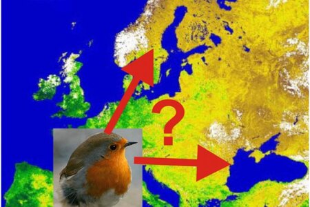

Migratory birds like European robins are able to use white or monochromatic light from the short-wavelength spectrum up to 565 nm to orient according to a magnetic compass system. Compass orientation is based on radical pair processes and is lateralized in favor of the right eye. For robins this implies a northbound direction towards Sweden during their spring migration. If, however, light with a long-wavelength component is given, the animals are no longer able to use their right-eye based compass system and depart to a 'fixed direction' response that is eastbound. Now a team of scientists from Frankfurt and Bochum universities demonstrated that these two visual orientation systems are differently organized with respect to hemispheric asymmetries. Robins were tested with either the right or the left eye covered or with both eyes uncovered for their orientation under different light conditions. With 502 nm turquoise light, the birds showed normal compass orientation, whereas they displayed an easterly 'fixed direction' response under a combination of 502 nm turquoise with 590 nm yellow light. Monocularly right-eyed birds with their left eye covered were oriented under turquoise in their northerly migratory direction, under turquoise-and-yellow towards east. The response of monocularly left-eyed birds differed: under turquoise light, they were disoriented, reflecting a lateralization of the magnetic compass system in favor of the right eye, whereas they continued to head eastward under turquoise-and-yellow light. Thus, 'fixed direction' responses are not lateralized. Hence und long-wavelength light the animals are no longer able to use their right-eye based compass system and depart to a non-lateralized 'fixed direction' response that is eastbound.

Executive functions such as set-shifting and maintenance are cognitive processes that rely on complex neurodevelopmental processes mediated by basal ganglia-prefrontal loops and the medial prefrontal cortex. To establish of basal ganglia-prefrontal loops, Reelin plays an important role in neurodevelopment. Although neurodevelopmental processes are mainly studied in animal models and in neuropsychiatric disorders, the underlying genetic basis for these processes under physiological conditions is poorly understood. We aimed to investigate the association between genetic variants of the Reelin (RELN) gene and cognitive set-shifting in healthy young individuals. The relationship between 12 selected single nucleotide polymorphisms (SNPs) of the RELN gene and cognitive set-shifting. We accounted for robust associations that were substantiated by subsequent haplotype analyses. The results suggest that variation in the REN gene affect processes of cognitive set-shifting and flexible rule adaptation and broadens the relevance of the RELN gene as a modulator for cognitive functions beyond neurological and/or psychiatric disorders.
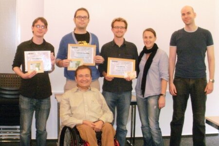

A delegation of our lab participated in the third autumn school of our DAAD-funded international PhD-program, taking part Sept. 19th-24th at the Hanse-Wissenschaftskolleg (HWK) in Delmenhorst. Doctoral fellows and their supervisors from Bochum, Izmir (Turkey) and Padua (Italy) came together for talks, vivid scientific discussions and poster sessions on both individual research projects and more general neuroscientific topics. Renowned scientists Josep Call from the MPI Leipzig, Germany, and Isabelle George from the CRNS in Rennes, France, completed the event as invited speakers.The doctoral fellows ran a competition for the best presentation and the best poster. For our lab, the autumn school was a success all down the line: We brought home all prizes. The picture below shows Sebastian Ocklenburg, the winner of the presentation prize, Felix Ströckens and Nils Kasties as winners of the poster prize, together with the organizational team, during the award ceremony.
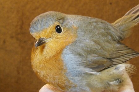

The magnetic compass orientation of birds is light dependent and is primarily mediated by the right eye. These findings make it likely that magnetic compass information is actually “seen” by the bird as a local change in its visual percept. The superiority of the right eye / left hemisphere in this process could then be a consequence of the superiority of the left hemisphere in discrimination tasks requiring object vision. Scientists from Frankfurt University and Biologists and Biopsychologists from Bochum have now conducted tests in the local geomagnetic field with European robins wearing goggles equipped with a clear and a frosted foil of equal optical translucence. Robins with a clear foil on the right eye and a frosted foil on the left eye oriented in the migratory direction as well as birds using both eyes. Birds with a frosted foil that blurred vision on the right eye and a clear foil on the left eye, in contrast, were disoriented. The publication in Current Biology is the first to show that avian magnetoreception requires, in addition to light, a nondegraded image formation along the projectional streams of the right retina. This suggests that the conversion of magnetic input into directional information requires processes that are also relevant for visual pattern recognition.

There is a growing interest in how pro-inflammatory cytokines modulate cognitive processes, especially since these molecules do not affect every brain area in a uniform way. The tumor necrosis factor alpha (TNF-alpha) for example exerts neuroprotective and neurodegenerative effects, depending on brain region. While some research suggests enhancing effects of the TNF-alpha gene (TNF-alpha -308G/A) on cognitive function, further research is needed to clarify the association between the TNF-alpha gene and specific areas of cognitive performance including their neurophysiological correlates. In this study we examined associations of the TNF-alpha -308G/A single nucleotide polymorphism (rs1800629) with attention and mental rotation performance in an event-related potential (ERP) study. The results show that carriers of the -308 A allele display elevated attentional processes (i.e. a stronger N1) as compared to the GG genotype group. Mental rotation performance varied across genotypes only when demands on mental rotation were high. Here, carriers of the -308 A allele performed better than the GG genotype group. This is paralleled by the neurophysiological data. The finding of enhanced attentional and mental rotation performance in A allele carriers supports recent findings that the A allele of this single nucleotide polymorphism (SNP) enhances cognitive performance on a general measure of cognitive processing speed.
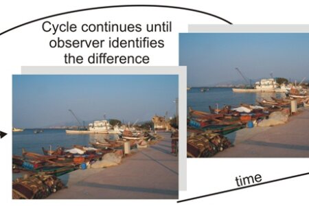

Do you see the difference between the two pictures? No? Then you are in good company. If the two pictures would simply alternate on a monitor, you would instantly see the difference. However, when a brief pause or a blank (like the grey partition in our example) is interposed between the two pictures, a person can sometimes gaze for minutes at the constantly switching two figures (and their interposed partitions) without detecting even very large changes in the scene. This phenomenon is called Change Blindness and it is to some extent still a mystery. Very likely, change blindness critically depends on attentional mechanisms. Visuo-spatial attention is lateralized towards the right hemisphere (left visual field). Wouldn’t we then expect change blindness to be more pronounced in the right visual field? In two successive experiments, psychologists from Izmir and Bochum tested this assumption and could prove it. Indeed, subjects could detect changes significantly faster in the left visual field, irrespective of their eye movements. Thus, even in cases when they started with a saccade to the right side of the picture, they were more likely to detect the difference on the left side of the picture.
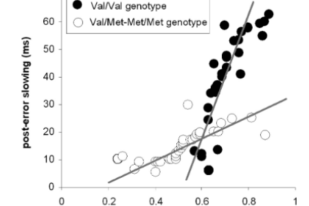

Behavioural adaptation depends on the recognition of response errors and processing of this error-information. Error processing is a specific cognitive function crucial for behavioural adaptation. Even though synchronization processes are important in information processing, its role and neurobiological foundation in behavioural adaptation are not understood. The brain-derived-neurotrophic-factor (BDNF) strongly modulates the establishment of neural connectivity that determines neural network dynamics and synchronization properties. Therefore altered synchronization processes may constitute a mechanism via which BDNF affects processes of error-induced behavioural adaptation. We investigate how variants of the BDNF gene regulate EEG-synchronization processes underlying error processing. Subjects (N=65) were genotyped for the functional BDNF Val66Met polymorphism (rs6265). We show that Val/Val genotype is associated with stronger error-specific phase-locking, compared to Met allele carriers. Post-error behavioural adaptation seems to be strongly dependent on these phase-locking processes and efficacy of EEG-phase-locking-behavioural coupling was genotype dependent. After correct responses, neurophysiological processes were not modulated by the polymorphism, underlining that BDNF becomes especially necessary in situations requiring behavioural adaptation. The results suggest that alterations in neural synchronization processes modulated by the genetic variants of BDNF Val66Met may be the mechanism by which cognitive functions are affected.


Pigeons use olfactory cues to navigate over unfamiliar areas when flying home. The left and the right hemispheres seem to play different roles in this process. In particular, the right olfactory bulb and left piriform cortex (the ‘cortical’-like area of olfactory representation) have been shown to be crucial for navigation. In a new study Biopsychologists from Bochum and Biologists from Pisa conducted a molecular imaging study in pigeons that had to home (or not) under different conditions. Imaging was done by the use of the immediate early gene ZENK that is activated when neurons are highly active. One group of pigeons was released from an unfamiliar site and had to fly home. On their way to the realease area they could smell and could use their olfactory sense when flying homeward. The second group was transported to the unfamiliar site (and could also smell everything), but was driven back without flying. The third group was released in front of the loft and had no trouble to find their way. In all groups, the nostrils of some pigeons were either occluded unilaterally or not. Released pigeons revealed the highest ZENK cell density in the olfactory bulb and the piriform cortex, indicating that very likely not smelling as such but the use of odours for homing from an unfamiliar site massively activates these structures. Only occlusion of the right olfactory bulb resulted in a decreased ZENK cell expression in the piriform cortex, whereas occlusion of the left nostril had no effect. This is the first study to reveal neuronal activation patterns in the olfactory system during homing. The data show that lateralized processing of olfactory cues is indeed involved in navigation over unfamiliar areas.


Es ist kaum zu glauben, aber Sabine Kesch hat gestern ihr 25jähriges Dienstjubiläum gefeiert. Seit so langer Zeit sorgt Sabine in der Biopsychologie, dass es den Tauben optimal geht. Sie fing zu einer Zeit an, in der dieser Lehrstuhl „Tierpsychologie“ hieß und der Chef Juan Delius. Später wurde Hans Markowitsch Professor des Lehrstuhls und benannte ihn in „Biopsychologie“ um. Noch später kam Onur Güntürkün. In all dieser Zeit hat Sabine die Tierpflege großartig organisiert und durchgeführt.
VIELEN DANK, SABINE!!!
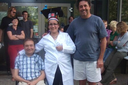

Josine Verhaal successfully fulfilled her defence on the 15th of July 2010 in the International Graduate School of Neuroscience (IGSN) and was awarded with a PhD. Josine gave a very nice overview of her work in which she showed how neurons in the entopallium code in a lateralized way for learned visual cues. She could show further how the interactions in an asymmetrically wired visual system are organized. One of her reviewers, Prof. Vern Bingman, came all the way from Ohio to this event. The picture shows her during the party after her defence along with Onur and Vern Bingman.
CONGRATULATIONS JOSINE!!!

Anxiety is often associated with impaired cognitive control that gets manifest because basal ganglia-prefrontal circuits are compromised, especially in anxiety disorders. The aim of this study was to investigate the effect of anxiety-related personality traits, such as anxiety sensitivity and trait anxiety, on event-related potentials of response inhibition in a standard Go/Nogo-paradigm that reflect processes of basal ganglia prefrontal circuits. We focused on the Nogo-N2 and Nogo-P3 components, which probably represent different sub-processes of response inhibition. The Nogo-N2 was mainly influenced by trait anxiety, while it was slightly affected by anxiety sensitivity. In contrast, the Nogo-P3 was significantly associated with anxiety sensitivity, but was less affected by trait anxiety. Thus, anxious subjects seem to maintain a higher level of cognitive control to prepare and to monitor the outcome of their actions, which is differentially reflected in Nogo-N2 and Nogo-P3 potentials. Our results show that anxiety-related personality traits modulate electrophysiological responses related to cognitive control processes and should be taken into consideration in studies investigating response inhibition. The results show that different types of anxiety seem to differentially modulate basal ganglia-prefrontal circuits.
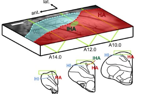

The pigeon is a widely established behavioral model of visual cognition, but the processes along its most basic visual pathways remain mostly unexplored. Now, Theoretical Neuroscientists and Biopsychologists from Bochum report the neuronal population dynamics of the visual Wulst, an assumed homolog of the mammalian primary visual cortex, captured for the first time with voltage-sensitive dye imaging. Responses to drifting gratings were characterized by focal emergence of activity that spread extensively across the entire Wulst, followed by rapid adaptation that was most effective in the surround. Using additional electrophysiological recordings, they found cells that prefer a variety of orientations. However, analysis of the imaged spatiotemporal activation patterns revealed no clustered orientation map-like arrangements as typically found in the primary visual cortices of many mammalian species. Instead, the vertical orientation was overrepresented, both in terms of the imaged population signal, as well as the number of neurons preferring the vertical orientation. Such enhanced selectivity for the vertical orientation may result from horizontal motion vectors that trigger adaptation to the extensive flow field input during flight and the fast head thrusts during walking. The findings suggest that, although the avian visual Wulst is homologous to the primary visual cortex in terms of its gross anatomical connectivity and topology, its detailed operation and internal organization is still shaped according to specific input characteristics.

Onur Güntürkün was accepted as a new member to the Wilhelm-Wundt Gesellschaft. The Wilhelm-Wundt Gesellschaft is a scientific society that has limited the maximal number of its scholars to 30. Its goal is the promotion of basic knowledge in the field of experimental psychology as well as the promotion of young scientists.

Pigeons are able to sense the magnetic field and use it to orient in space. However, a proof of magnetic compass learning by pigeons under laboratory conditions has been attempted for decades, but all experiments have failed so far. Now biopsychologists from Bochum and animal behavioral scientists from Frankfurt University aimed to test whether pigeons can learn magnetic compass directions in an operant chamber if magnetic cues are presented as true spatial cues. Experimental sessions were carried out in the local geomagnetic field and in magnetic fields with matched total intensity and inclination, but different directions generated with Helmholtz-coils. Birds demonstrated successful learning with a performance level comparable to that in learning studies with magnetic anomalies. Surprisingly, the most successful magnetic field learners in the lab were subsequently those that first took a detour in the field before flying to their loft. Did they first explore the territory before embarking on their voyage? In any way, these findings represent the first evidence for operant magnetic compass learning in pigeons and also provide a link between behavioural data from the field and the laboratory.
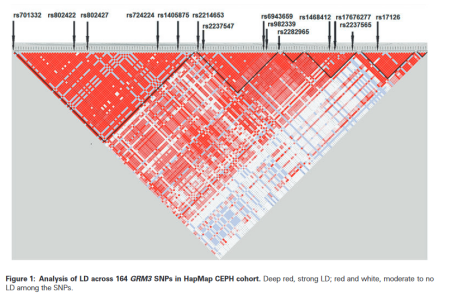

The ability to adapt behaviour in a flexible way is a crucial cognitive function. Set-shifting and maintenance are complex cognitive processes, which are often impaired in several neuropsychiatric and neurodegenerative diseases. The genetic basis of these processes is poorly understood. We aimed to investigate the association between genetic variants of the metabotropic glutamate receptor 3 (GRM3) and cognitive set-shifting in healthy individuals. The relationship between 14 selected single nucleotide polymorphisms (SNPs) of the GRM3 gene and cognitive set-shifting as measured by perseverative errors using the modified card sorting test (MCST). Results show that SNP rs17676277 is related to the performance on the MCST. Subjects with the TT genotype showed significantly less perseverative errors as compared with the AA (P = 0.025) and AT (P = 0.0005) and combined AA/AT genotypes (P = 0.0005). Haplotype analyses suggest the involvement of various SNPs of the GRM3 gene in perseverative error processing in a dominant model of inheritance. The findings strongly suggest that the genetic variation (rs17676277 and three haplotypes) in the metabotropic GRM3 is related to cognitive setshifting in healthy individuals independent of working memory.
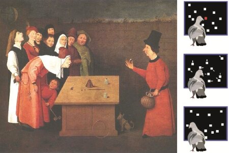

For sure you know how difficult it is to track the correct shell in a shell game (German: “Hütchenspiel”). We have to fully attend to the correct object to not lose track of it. Selective attention is a crucial component of all sensory processing. Biopsychologists from Bochum tested the role of dopamine in attentional selection and in the maintenance of attention in pigeons. The birds were trained on a moving-dot paradigm comparable to the shell game and had to select a target among distractors and maintain attention to the target. Target and distractors consisted of white dots, moving at random on a touch-screen. Here you see a historic picture of the shell game along with a cartoon of three sequences of the experiment conducted with pigeons. In the pigeon-version of the shell game, the demand on attention was modulated by varying the number of distractors and the duration of motion. Both manipulations affected performance equally. In the next step, the contribution of dopamine to attention was investigated. Intracranial injections of D1-antagonist before testing led to decrements in performance that equally affected trials with different attentional demand. This drop in performance could not be attributed to altered motivation or motor performance. The conclusion was that dopamine has a critical role in attention. It is involved in the selection of targets for attention and in the stabilization of attention against interference. This is comparable to the role dopamine plays in working memory and argues for similar mechanisms underlying selective attention and working memory. For a video of the experiment, see below:
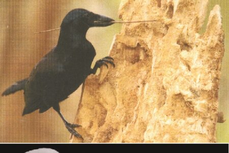

Animals with a high rate of innovative and associative-based behaviour usually have large brains. New Caledonian (NC) crows stand out due to their tool manufacture, their generalized problem-solving abilities and an extremely high degree of encephalization. It is generally assumed that this increased brain size is due to the ability to process, associate and memorize diverse stimuli, thereby enhancing the propensity to invent new and complex behaviours in adaptive ways. However, this premise lacks firm empirical support since encephalization could also result from an increase of only perceptual and/or motor areas. Scientists from the Bochum Biopsychology lab, the University of Düsseldorf and the University of Auckland (New Zealand) now compared the brain structures of NC crows with those of other advanced birds. The brains of NC crows were characterized by a relatively large association and motor-learning areas. This supports the hypothesis that the evolution of innovative or complex behaviour requires a brain composition that increases the ability to associate and memorize diverse stimuli in order to execute complex motor output. Since apes show a similar correlation of cerebral growth and cognitive abilities, the evolution of advanced cognitive skills appears to have evolved independently in birds and mammals but with a similar neural orchestration.
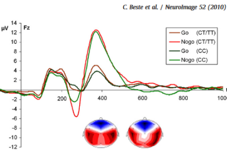

The basal ganglia are connected to prefrontal cortical areas via different functional loops. These loops do not only differ with respect to their cognitive functions the mediate, but also on a neurochemical level. In the current study we ask, how these differences on a neurobiochemical level may affect cognitive processes mediated by these loops. One function of the anterior-cingulate loop is error processing. One function of the orbito-frontal loop is response inhibition. Combining a genetic approach with event-related potential (ERP) measurements of response inhibition (OFC-loop function) and error processing (ACC-loop function), we provide robust results showing a selective modulation of response inhibition processes by the GRIN2B C2664T polymorphism at the behavioural and neurophysiological level. Since error processing functions were not affected, the results suggest for differential influences of the GRIN2B C2664T polymorphism on response inhibition and error processing functions. The results provide first insight into cognitive-neurophysiological effects of the GRIN2B C2664T polymorphism. The dissociation obtained may be due to a differential importance of N-methyl-d-aspartate receptors for glutamatergic neural transmission in different striatal compartments (matrix and striosomes). We provide a model on this that may be a target for future research.
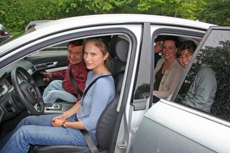

The stereotype of women’s limited parking skills is deeply anchored in modern culture. When entering the items ‘women’ and ‘parking’ in one of the biggest search engines of the World Wide Web, more than 80.000.000 results are obtained. As parking is a complex, spatial task, and a large body of literature proves the existence of sex differences in spatial cognition in favour of men, it is possible that the prejudice addressing women’s poor parking skills has a scientific background. To test this possibility, we investigated parking performance of 17 driving beginners and 48 more experienced drivers in an area of a car park that was closed off for the public. Subjects conducted three different types of manoeuvres (forward, backward, parallel), which were analysed for speed and accuracy. We found that men park more accurately and especially faster than women. On average, males were 42 seconds faster and 3 % more accurate than women. Although overall performance was better in experienced drivers, the sex difference remained. Performance was related to spatial skills and self-assessment in driving beginners, but only to self-assessment in more experienced drivers. As male subjects outperformed females in the mental rotation test for spatial skills and achieved higher scores in the self-assessment questionnaire, we assume that the sex difference in parking is due to these two variables. We assume that – due to differential feedback – self-assessment incrementally replaces the controlling influence of spatial skills, as parking is trained with increasing experience. Additionally, the prevalence of the prejudice about women’s parking skills could constitute a stereotype threat that additionally decreases women’s performance. Our results suggest that sex differences in spatial cognition found in laboratory experiments persist in real-life situations. However, real-life spatial cognition is also influenced by socio-psychological factors, which modulate the biological causes of sex differences.

The movement of animals through space filled with various objects requires the interaction between neuronal mechanisms specialized for processing local object motion and those specialized for processing optic flow generated by self-motion of the animal. In the avian brain, visual nuclei in the tectofugal pathway are primarily involved in the detection of object motion. By contrast, the nucleus of the basal optic root (nBOR) and the pretectal nucleus lentiformis mesencephali (nLM) are dedicated to the analysis of optic flow. But little is known about how these two systems interact. Using single-unit recording in the entopallium of the tectofugal pathway, we show that some neurons appeared to be integrating visual information of looming objects and whole field motion simulating optic flow. They specifically responded to looming objects, but their looming responses were modulated by optic flow. Optic flow in the nasotemporal direction, typically produced by the forward movement of the bird, only mildly inhibited the looming responses. Furthermore, these neurons started firing later than when the looming object was presented against a stationary background. However, optic flow in other directions, especially the temporonasal direction, strongly inhibited their looming responses. Previous studies have implicated looming-sensitive neurons in predator avoidance behavior and these results suggest that a bird in motion may need less time to initiate an avoidance response to an approaching object.
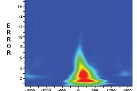

Behavioural adaptation and cognitive control are crucial for goal-reaching behaviour. Common sense suggests that errors are an important source of information in the regulation of such processes. Much effort is undertaken to understand the neurobiological mechanisms underlying these processes. Several lines of research suggest that the serotonergic system may be of special interest in this respect.
This study investigates the dependence of response monitoring and error detection on genetic influences modulating the serotonergic system, which was done using the event-related potentials (ERPs) after error (Ne/ERN) and correct trials (Nc/CRN). To induce a sufficient amount of errors, a standard flanker task was used. The subjects (N = 94) were genotyped for the functional 5-HT1A C(-1019)G polymorphism. In order to investigate neural processes that are specific for error monitoring, time-frequency analyses (wavelet-analyses) were applied. The results show that the 5-HT1A C(-1019)G polymorphism specifically modulates error detection. Neurophysiological modulations on error detection were paralleled by a similar modulation of response slowing after an error, reflecting the behavioral adaptation. The 5-HT1A -1019 CC genotype group showed a larger Ne and stronger post-error slowing than the CG and GG genotype groups. More general processes of performance monitoring, as reflected in the Nc/CRN, were not affected. The finding that error-specific processes, but not general response monitoring processes, are modulated by the 5-HT1A C(-1019)G polymorphism is underlined by a wavelet analysis. In summary, the results suggest a specific effect of the 5-HT1A C(-1019)G polymorphism on error monitoring processes and suggest a neurobiological dissociation between processes of error monitoring and general response monitoring at the level of the serotonin 1A receptor system.
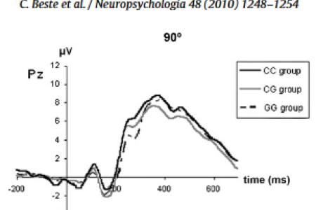

Numerous lines of research indicate that attentional processes, working memory and saccadic processes are highly interrelated. Even though this relation has often been postulated in literature, the neurobiological mechanism underlying this interrelation, are not understood. In the current study, we examine the relation between these processes with respect to their cognitive-neurophysiological and neurobiological background by means of event-related potentials (ERPs) in a sample of N = 72 healthy probands characterized for the functional serotonin receptor 1 A (5-HT1A) C(−1019)G polymorphism. The results support a close interrelation between working memory, attentional and saccadic processes. Yet, these processes are differentially modulated by the 5-HT1A C(−1019)G polymorphism. The 5-HT1A C(−1019)G polymorphism primarily affects attentional processing, whereas processes related to the mental rotation of an object are independent of 5-HT1A genetic variation. It is shown that an increasing number of −1019 G alleles leads to a differential reduction of the N1 above the left and right hemisphere and hence bottom-up attentional processing. In the way increasing numbers of −1019 G alleles lead to a reduction of attentional processes, saccadic activity increases as a similar function of the number of −1019 G alleles. This increase in activity occurs parallel in time to the process of mental rotation. It is hypothesized that decreased attentional processes, dependent on different 5-HT1A C(−1019)G genotypes, may cause parietal networks to increase saccadic activity in order to perform mental rotation. The results support theories of highly interlinked attention, working memory and eye-movement systems.
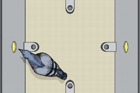

How does it feel to sense the magnetic field? How is it to actually "see" where North is? We humans do not know how it feels to sense the magnetic compass but we can study the mechanisms of this system in birds. Previous studies had shown that birds can "visualize" the magnetic compass directions and, in addition, can sense the strength of the local magnetic field. However, all attempts to actually train birds to learn a compass direction had failed. A new study by scientists from Frankfurt, Düsseldorf and the Biopsychology Department in Bochum now reports the first successful study on learning the magnetic compass direction. Pigeons were trained in an operant chamber with magnetic compass directions as true spatial cues. Experimental sessions were carried out in the local geomagnetic field and in magnetic fields with matched total intensity and inclination, but divergent directions generated with Helmholtz-coils. The animals successfully learned to peck those keys that were in the direction of the correct compass direction. Subsequently, the birds were tested in a true outdoor homing task. Astonishingly, the more successful individuals of the Skinner-Box learning experiment had worse initial orientations at unfamiliar, but not at familiar sites. The authors speculate that the better learners might have more self confidence to first explore unknown locations before taking the way back home. Future studies will show if this interpretation is correct.
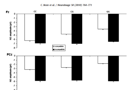

Response selection and control are supposed to reflect important basal ganglia functions. Recently, we showed that the dopaminergic system may be especially important for response selection in compatible, but not in incompatible stimulus-response (S-R) relations. Research indicates that the dopaminergic system is influenced by the serotonergic system, but little is known about the involvement of the serotonergic system in response selection. Analyzing event-related potentials (ERPs) we show the 5-HT1A C(-1019)G polymorphism modulating response-related processes, but only in compatible S-R relations. This modulation was a function of the number of -1019 G alleles. Decreasing numbers of -1019 G alleles were stepwise related to increases in response selection efforts. The functional effect of the 5-HT1A C(-1019)G polymorphism has previously been shown to be specific for serotonergic 1 A autoreceptors of serotonergic neurons in the dorsal raphe nucleus (DRN). Due to this close relation of genotype effects to neuroanatomically dissociable structures, the results suggest that DRN serotonin 1 A autoreceptors are important for response selection. The results extend previous findings on the dopaminergic system to the serotonergic system. The examined functions are precisely regulated on a neuronal level, since neurophysiological and behavioural effects are driven in an allele-dose fashion. Because of this, the results are of importance for future clinical applications.
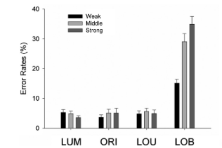

The way our brain selects relevant information and discards irrelevant one is a highly competitive process. The ability to notice relevant visual information has been assumed to be determined both by the relative salience of relevant information compared with distracters within a given display and by voluntary allocation of attention toward intended goals. A dominance of either of these two mechanisms in stimulus processing has been claimed by different theories. A central question in this context is to what degree and how task irrelevant signals can influence processing of target information. This question was examined in this study using event-related potentials (ERPs). Using parametrical manipulations of the saliency of distracting stimuli we found that the amount of saliency was predictive for the proportion of detected information when relevant and irrelevant information were spatially separated but not when they overlapped. Weighting and competition of incoming signals was reflected in the amplitude of the N1pc component of the event-related potential. Initial orientation of attention toward the irrelevant element had to be followed by a reallocation process, reflected in an N2pc. The control of conflicting information additionally evoked a fronto-central N2 that varied with the amount of competition induced. Thus the data support models that assume that attention is a dynamic interplay of bottom-up and top-down processes that may be mediated via a common dynamic neural network.
Wascher, E., & Beste, C., Tuning perceptual competition, J Neurophysiol, 2010, 103: 1057-1065.


Tobias Ohmann successfully fulfilled his defence on the 9th of February 2010 in the Faculty of Psychology and was awarded with a Dr. rer. nat. Tobias truly impressed the committee and the spectators (the room was filled to the last place) with a brilliant overview of his work and a smooth and highly informed discussion. In his thesis, Tobias had studied the behavioural neuroscience of time – a highly elusive property of life. His experiments reached from waiting for reward or punishment up to episodic-like recall of the last time-of-meal and incorporated highly ingenious behavioural approaches.
CONGRATULATIONS TOBIAS!!!
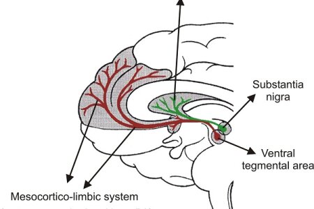

Response inhibition is a component of executive functions, which can be divided into distinct subprocesses by means of event-related potentials (ERPs). These subprocesses are (pre)-motor inhibition and inhibition monitoring, which are probably reflected by distinct ERP-components. We asked, if these subprocesses may depend on distinct basal ganglia subsystems. We examined response inhibition processes in an extended sample of young and elderly subjects, patients with Parkinson’s disease (PD) and Huntington’ disease (HD). This combination of groups also allow us to study whether, and to what degree, pathological basal ganglia changes and healthy aging have similar and/or different effects on these processes. Indeed subprocesses of response inhibition are differentially modulated by distinct basal ganglia circuits. Processes related to (pre)-motor inhibition appear to be modulated by the nigrostriatal system, and are sensitive to aging and age-related basal ganglia diseases (i.e. PD). Parkinson’s disease induces additive effects of aging and pathology. In contrast, inhibition monitoring is most likely modulated by the mesocortico-limbic dopamine system. These processes are equally affected in healthy aging and both basal ganglia diseases (i.e. PD, HD).
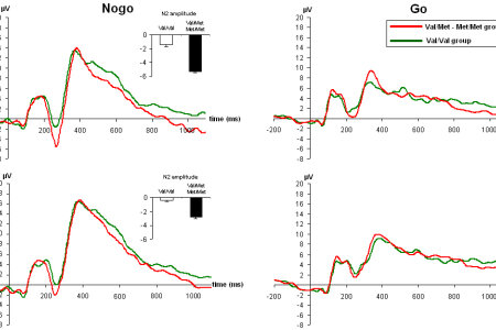

Response inhibition is a basic executive function which is dysfunctional in various basal ganglia diseases. The brain-derived-neurotrophic-factor (BDNF) plays an important pathophysiological role in these diseases. In the current study we examined the functional relevance of the BDNF val66met polymorphism for response inhibition processes in 57 healthy human subjects using event-related potentials (ERPs), which likely reflect different aspects of inhibition. The results show that the BDNF val66met polymorphism selectively modulates the pre-motor subprocesses of response inhibition. Response inhibition was better in the val/met-met/met group, since this group committed fewer false alarms, and their Nogo-N2 was larger, compared to the val/val group. This is the first study showing that met alleles of the BDNF val66met polymorphism confer an advantage for a specific cognitive function. We propose a neuronal model how this advantage gets manifest on a neuronal level.

Peripheral and central mechanisms of pain from incision differs from inflammatory or chronic pain. It is expected that brain activation patterns differ with pain type but have not been studied in humans. In this study, the activation of different brain areas after an experimental surgical incision was assessed by functional magnetic resonance imaging, and the pathophysiological role of distinct brain activation patterns for pain perception after incision was analyzed. Functional magnetic resonance imaging analysis showed a distinct temporal profile of activity within specific brain regions during and after the injury. Lateralization (predominantly contralateral to the incision) and increased brain activity of the somatosensory cortex, frontal cortex, and limbic system were observed in subjects after incision, when compared with individuals receiving sham procedure. Peak brain activation occurred about 2 min after incision and decreased subsequently. Basal ganglia structures and especially the caudate nucleus were activated shortly after incision. Lateron activity persists in neocortical structures, but was absent in basal ganglia structures. A distinct correlation between evoked pain ratings and brain activity was observed for the anterior cingulate cortex, insular cortex, thalamus, frontal cortex, and somatosensory cortex. These findings show different and distinct cortical and subcortical activation patterns over a relevant time period after incision. Pain sensitivity hereby has an influence on the activity profile. This may have important implications for encoding ongoing pain after a tissue injury, for example, resting pain in postoperative patients.
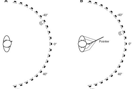

Several studies have shown that handedness has an impact on visual spatial abilities. However, the relationship of handedness and spatial processing in the auditory modality has remained largely unclear. To investigate this relationship, scientist from Durham University (England), the Ifado in Dortmund and the Ruhr-University of Bochum assessed auditory space perception in left- and right-handers in a dark, anechoic, and sound-proof room. Interestingly, participants showed a bias in sound localization that was to the side contralateral to the preferred hand. This partially parallels findings in the visual modality as left-handers typically have a more rightward bias in visual line bisection compared with right-handers. Despite the differences in neural processing of auditory and visual spatial information these findings show similar effects of lateral preference on auditory and visual spatial perception. This suggests that supramodal neural processes are involved in the mechanisms generating laterality in space perception.

Most theories of human handedness assume that it is completely determined by genetic factors. However, since there is a large body of evidence for an influence of non-genetic factors on asymmetries in other vertebrates, biopsychologist from Bochum tested the hypothesis that human handedness may also result from a combination of genetic and experience-driven factors. In order to do so, the research team analysed sidedness in children suffering from congenital muscular torticollis. These children display a permanently tilted asymmetric head posture to the left or to the right in combination with a contralateral rotation of face and chin, which could lead to an increased visual experience of the hand contralateral to the head-tilt. Relative to controls, torticollis children had a higher probability of right- or left-handedness when having a head-tilt to the opposite side. Thus, an increased visual control of the hand during early childhood seems to modulate handedness. These findings not only show that human handedness is affected by early lateralised visual experience but also speak in favour of a combined gene-environment model for its development.
![]()
Cicero (106-43 B.C.) is quoted as saying, 'Ut imago est animi voltus sic indices oculi' (the face is a picture of the mind as the eyes are its interpreter). Today we use this original quote in the slightly abbreviated form as “the eyes are the window to the soul”. Indeed, countless eye-tracking studies have uncovered hidden intentions and hot spots of attention by observing the eye movements of human volunteers. Unfortunately, bird researchers were until recently excluded from this rich source of knowledge since it is impossible to comparably track eye movements of birds. Now, scientists from the Biopsychology laboratory in Bochum invented a fast way to track the peck locations of pigeons while these animals discriminated hundreds of photographs that contained humans or just landscapes and other objects. Peck location tracking is an indirect way to trace the foci of attention of pigeons. Once this method was established, the scientists were up to a surprise: The birds specifically pecked onto the humans in the pictures. Thus, they attended to the critical portion of the pictures and aimed at these areas. But even more than that: the animals focused on the heads of humans. When these heads were cut out, discrimination success of the birds dropped significantly. So, reading a pigeon’s mind reveals that for them it is our face that makes us human.
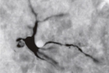

Tetrodotoxin, the poison of the buffer fish, is often used by neuroscientists to selectively block specific brain regions. In spite of this abundant use, little was known about the temporal and spatial decay of TTX injected in brain tissue. Scientists of the Biopsychology group have now been able to develop an immunohistochemical protocol to make injected TTX visible on brain slices. With this approach they could show that the diffusion of TTX one hour after injection to pigeons' entopallium was 3 mm in all directions. Furthermore, contrary to the often assumed decay period of maximally 24 h, they showed that only after 48 hours TTX had completely cleared from the brain tissue. With this new approach the spread of injected TTX can easily be visualized and therefore it makes the blocking of specific brain regions more reliable.

It seems to be so easy to recognize a person on a photograph. In fact, it’s quite difficult: First of all, we have to transform a 3D-human to a 2D-image. We also have to accept that the absolute size of the two objects is vastly different, that the human moves around while the picture is static, etc. Despite these and many more problems, we easily generalize from humans to pictures and vice versa. But are other animals also able to do it? This is an important question since hundreds of publications with pigeons rely on the implicit assumption that photographs do represent real objects for these birds. Biopsychologists from Bochum, from the Max-Planck-Institute for Biological Cybernetics in Tübingen and from Mugla University (Turkey) tested the ability of pigeons to perform the transfer from 3D-individual humans to 2D-pictures of these individuals. Sixteen birds were first trained to identify individual, real-life humans. It turned out that this discrimination depended primarily on visual cues from the heads of the persons. Subsequently, the pigeons were shown photographs of these individuals to test for transfer to a two dimensional representation. Successful identification of a three-dimensional person did not facilitate learning of the corresponding photographs. These results show that the cross-recognition of individual real-life humans and their photographs in pigeons is far more limited than previously assumed.
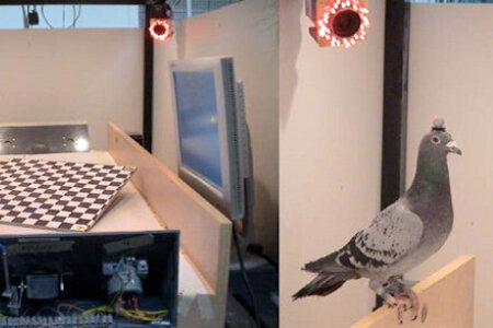

Have you ever watched a pigeon walking? The head constantly seems to thrust foreward and backward while the rest of body behaves as if being a wind up toy. Scientists know since long that the fore-and-back oscillation of the head is just an illusion: In reality the head quickly thrusts foreward but subsequently stands still while the body moves. Then the head thrusts foreward again, etc. Thus, there is no backward movement of the head. But why is the movement of the head different from the rest of the body? Well, here comes a clever explanation: If the head would move constantly together with the body, the retinal image would be blurred due to movement artefacts. So, a quick foreward thrust (during which the animal should be blind mostly), followed by hold period should be an optimal solution. This explanation was so compelling that it was accepted by almost everybody for decades. Now scientists from Spain, Canada, and Biopsychologists from the Ruhr-University have killed this theory (requiescat in pace as the Romans would say). With a true high-tech approach they managed to flash patterns onto monitors while the pigeons were either just thrusting their head foreward, or were just in their hold phase. To do so, head movements were tracked by means of an optical motion capture system consisting of high-speed cameras which provided the three-dimensional position of a marker mounted on the head of the animal in real time. The job of the pigeons was simple: Tell me which pattern was shown to you. The results were also simple: The pigeons were equally able to perfectly perform the discrimination both in the thrust and in the hold phase. Thus, head-bobbing can not be explained by a temporary blindness during the movement time of the head. Now that the temporary-blindness-theory is dead, what then is the explanation for head bobbing? Well, more research is needed…


You read this paragraph by mostly using resources of your left hemisphere. But is the asymmetry of your brain realized at the neuronal level and how does it emerge during your lifetime? The pigeon’s visual system is an excellent model to find answers for these questions. Now, Biopsychologists from Bochum have published a review on 20 years of asymmetry research in this model. They describe how lateralized visual stimulation induces structural asymmetries within the brain of the embryo during a critical time window, and how these early asymmetrical signals shape the nervous system into a lateralized structure. Interhemispheric control mechanisms seem to stabilize these induced asymmetries. The functional left-right differences are then established due to the interplay between bottom-up and top-down mechanisms that generate a lateralized, hemispheric-specific visual analysis.

Lars Dittrich successfully graduated on the 29th of October in the IGSN and was awarded with a PhD of Neuroscience. Lars impressed the committee so much that they unanimously decided to grade his thesis and defence with a summa cum laude – a rare and very prestigious event. Lars analyzed how pigeons learn a complex categorization task in which the animals have to come up with a strategy to discriminate humans from non-humans on pictures. In addition he introduced novel ways to "read a pigeons mind" with behavioural and anatomical means. Subsequent to the party, Lars' students gave him a shisha as present. As shown below, the new device was directly put into widespread use.
CONGRATULATIONS LARS!!!


Maik Stüttgen received a teaching award from the students of the Graduate School for Neural and Behavioural Sciences. During the winter term 2009, he lectured on 'Essential Statistics' for students of neuroscience at the University of Tübingen. The curriculum covered a range of topics including probability theory, correlation & regression, null-hypothesis statistical testing, effect sizes and confidence intervals.
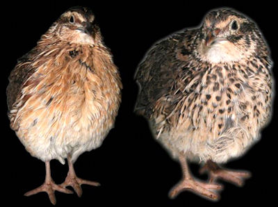

Japanese quail are an excellent model for studies on sexual behaviour. They very quickly learn to approach quails of the opposite sex when being reinforced with access to copulation. Previous studies had shown that quails tested with their left eye performed better than the ones tested with their right eye when working for sex. This right hemispheric dominance for instinctual and affective behaviour is typical for vertebrates, incl. humans. Now scientists from the Bochum Biopsychology lab and two universities in Izmir (Turkey) aimed to see if this hemispheric dominance can be modified by asymmetrical light input in early life. To this end they closed the right or the left eyes of the 2 day-old quail for 70 days and tested the subjects in the same set up. Indeed, asymmetry of behaviour had disappeared. Thus, brain asymmetries can be altered with asymmetrical sensory stimulation in early life. However, the scientist were up for a surprise when they removed the eye cap from the closed eye, switched its position to the previously open eye and retested the birds. Suddenly, the old right hemispheric dominancy emerged again. So, the old left-right differences had survived in the deprived hemisphere, showing that lateralized circuits are retained to some extent, even after major changes on the undeprived side.
In January 2008 the seminar "Das menschliche Gehirn - ein Mal- und Bastelkurs" (The human brain - a painting- and craftsmanship course) of the Biopsychology department was awarded on the conference "Kompetenzorientiert lehren und lernen an der RUB" for exemplary teaching.
As a result this film was made to show the concept and the realization of the seminar to a broad community.
If you enjoyed it please distribute the web location to people who might be interested in it.