![]()
![]()
![]()
![]()
2012-11-22

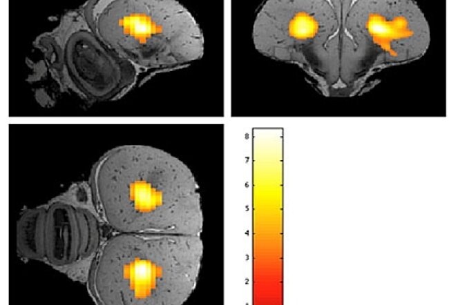
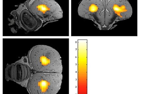
Functional MRI (fMRI) is widespread in humans but still rare in animal research. If being applied, nearly always anaesthetized animals are used. Only a few articles present results obtained in awake rodents. With advent of the "cognitive bird revolution", awake bird fMRI is increasingly relevant. Bioimaging experts from Antwerp and Biopsychologists from Bochum now conducted the first awake bird imaging study with highly habituated and head fixed pigeons. Both traditional fMRI and resting state (rsfMRI) were applied. In addition, this is the first time functional connectivity measurements were performed in a non-mammalian species. Since the visual system of pigeons is a well-known model for brain asymmetry, the focus of the study was on the neural substrate of the visual system. For fMRI a visual stimulus was used and functional connectivity measurements were done with the entopallium as a seed region. Interestingly in awake pigeons the left E was significantly functionally connected to the right E. Moreover connectivity maps for a seed region in both hemispheres resulted in a stronger bilateral connectivity starting from left E. These results could be used as a starting point for further imaging studies in awake birds and also provide a new window into the analysis of hemispheric dominance in the pigeon.
Functional MRI (fMRI) is widespread in humans but still rare in animal research. If being applied, nearly always anaesthetized animals are used. Only a few articles present results obtained in awake rodents. With advent of the "cognitive bird revolution", awake bird fMRI is increasingly relevant. Bioimaging experts from Antwerp and Biopsychologists from Bochum now conducted the first awake bird imaging study with highly habituated and head fixed pigeons. Both traditional fMRI and resting state (rsfMRI) were applied. In addition, this is the first time functional connectivity measurements were performed in a non-mammalian species. Since the visual system of pigeons is a well-known model for brain asymmetry, the focus of the study was on the neural substrate of the visual system. For fMRI a visual stimulus was used and functional connectivity measurements were done with the entopallium as a seed region. Interestingly in awake pigeons the left E was significantly functionally connected to the right E. Moreover connectivity maps for a seed region in both hemispheres resulted in a stronger bilateral connectivity starting from left E. These results could be used as a starting point for further imaging studies in awake birds and also provide a new window into the analysis of hemispheric dominance in the pigeon.
Copyright © BioPsy 2023
Last update: Oct 03, 2023