![]()
![]()
![]()
![]()
2016-05-24

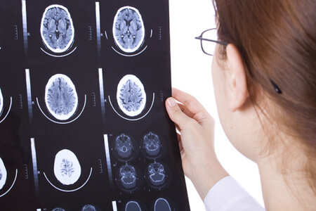
Imagine that you undergo training as a neurologist and have to group MR-images into those with and those without a tumor. Once you spot a tumor, you could simple memorize this image, and could decide on future images based on the similarity to the first tumor you saw. This way to categorize tumors is called “exemplar”-based categorization. But you could also derive an abstraction from your first tumor (its general properties) and use this summary representation for your future diagnosis. This strategy would be a “prototype”-based categorization. Subjects tested in such tasks often show different learning curves for prototype and exception stimuli. They first learn based on prototypes and then learn odd stimuli based on exemplar strategies. Neuro- and biopsychologists from Bochum now examined the contributions of different brain structures to prototype and exemplar-based category learning using functional magnetic resonance imaging (fMRI). Indeed, their subjects showed an initially superior performance for prototypes and then switched to an exemplar-based categorization for exceptions in the later learning phases. Analysis of the functional imaging data revealed that the left fusiform gyrus was involved in processing of prototypes while an activation of the right hippocampus correlated with processing of exceptions. Thus, successful prototype- and exemplar-based category learning is associated with activations of complementary neural substrates that constitute object-based processes of the ventral visual stream and their interaction with unique-cue representations, possibly based on sparse coding within the hippocampus.
Imagine that you undergo training as a neurologist and have to group MR-images into those with and those without a tumor. Once you spot a tumor, you could simple memorize this image, and could decide on future images based on the similarity to the first tumor you saw. This way to categorize tumors is called “exemplar”-based categorization. But you could also derive an abstraction from your first tumor (its general properties) and use this summary representation for your future diagnosis. This strategy would be a “prototype”-based categorization. Subjects tested in such tasks often show different learning curves for prototype and exception stimuli. They first learn based on prototypes and then learn odd stimuli based on exemplar strategies. Neuro- and biopsychologists from Bochum now examined the contributions of different brain structures to prototype and exemplar-based category learning using functional magnetic resonance imaging (fMRI). Indeed, their subjects showed an initially superior performance for prototypes and then switched to an exemplar-based categorization for exceptions in the later learning phases. Analysis of the functional imaging data revealed that the left fusiform gyrus was involved in processing of prototypes while an activation of the right hippocampus correlated with processing of exceptions. Thus, successful prototype- and exemplar-based category learning is associated with activations of complementary neural substrates that constitute object-based processes of the ventral visual stream and their interaction with unique-cue representations, possibly based on sparse coding within the hippocampus.
Copyright © BioPsy 2023
Last update: Sep 28, 2023