![]()
![]()
![]()
![]()
2018-04-25

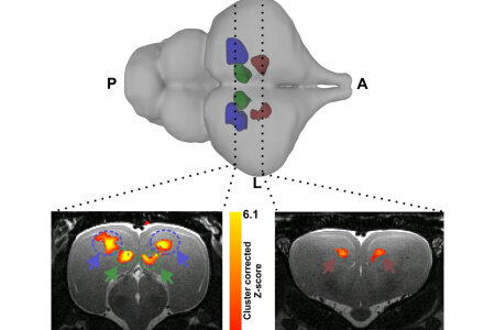
The phylogenetic position of crocodilians in relation to birds and mammals make them an interesting animal model to investigate the evolution of sensory systems in vertebrates. An appropriate, non-invasive method that can be utilized in such studies is fMRI. An international group of researchers from the Biopsychology Department and the School of Anatomical Science at the University of the Witwatersrand (South Africa) now employed fMRI, never previously tested in poikilotherms, to investigate crocodilian telencephalic sensory processing. Juvenile Crocodylus niloticus were placed in a 7T MRI scanner and BOLD signal changes were recorded during the presentation of visual (flickering light at 2-8 Hz) and auditory (simple: chords centered around 1000 or 3000 Hz, and complex: classic music) stimuli. Our findings confirm that the majority of activated regions to both visual and auditory stimuli parallel what has been described for mammalian and avian sensory pathways, indicating conserved sensory processing principles among amniotes. The application of fMRI to ectothermic vertebrates provides an avenue for the application of this method to future functionally related brain research in a broader spectrum of vertebrate species.
The phylogenetic position of crocodilians in relation to birds and mammals make them an interesting animal model to investigate the evolution of sensory systems in vertebrates. An appropriate, non-invasive method that can be utilized in such studies is fMRI. An international group of researchers from the Biopsychology Department and the School of Anatomical Science at the University of the Witwatersrand (South Africa) now employed fMRI, never previously tested in poikilotherms, to investigate crocodilian telencephalic sensory processing. Juvenile Crocodylus niloticus were placed in a 7T MRI scanner and BOLD signal changes were recorded during the presentation of visual (flickering light at 2-8 Hz) and auditory (simple: chords centered around 1000 or 3000 Hz, and complex: classic music) stimuli. Our findings confirm that the majority of activated regions to both visual and auditory stimuli parallel what has been described for mammalian and avian sensory pathways, indicating conserved sensory processing principles among amniotes. The application of fMRI to ectothermic vertebrates provides an avenue for the application of this method to future functionally related brain research in a broader spectrum of vertebrate species.
Copyright © BioPsy 2023
Last update: Sep 28, 2023