![]()
![]()
![]()
![]()
2023-07-28

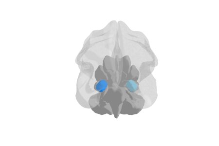
The amygdala is an evolutionarily conserved core structure in emotion processing and one of the key regions of interest in affective neuroscience. In a new review researcher from the biopsychology lab highlighted recent findings on amygdala neuroimaging.
They focused on an important problem: Results of neuroimaging studies on the amygdala are often heterogeneous since it is composed of functionally and neuroanatomically distinct subnuclei. Fortunately, ultra-high-field imaging offers more accurate functional and structural representations of subnuclei and their connectivity. Most clinical studies using ultra-high-field imaging focused on major depression, suggesting either overall rightward amygdala atrophy or distinct bilateral patterns of subnuclear atrophy and hypertrophy. Connectivity analyses identified widespread networks for learning and memory, stimulus processing, cognition, and social processes, thereby provide evidence for distinct roles of different amygdala subnuclei. The researchers propose theoretical and methodological considerations that will help future ultra-high-field imaging to disentangle the ambiguity of the amygdala’s function, structure, connectivity, and clinical relevance.
Kirstein CF, Güntürkün O, Ocklenburg S. (2023). Ultra-high field imaging of the amygdala - A narrative review. Neurosci Biobehav Rev, 152, 105245.
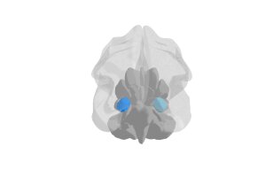
Copyright © BioPsy 2023
Last update: Jul 28, 2023